BOTANY

CHAPTERS 1-9
BOOKLET-1
Contents: Page No.
Chapter 1 Cell- The Unit of Life Part 1 1-17
Chapter 2 Cell- The Unit of Life Part 2 18-36
Chapter 3 Cell- The Unit of Life Part 3 37-43
Chapter 4 Cell- The Unit of Life Part 4 44-54
Chapter 5 Biomolecules 55-68
Chapter 6 Cell Cycle and Cell Division 69-82
Chapter 7 Biological Classification Part 1 83-102
Chapter 8 Biological Classification Part 2 103-108
Chapter 9 Biological Classification Part 3 109-122
| Cell as a unit of life. |
(1) Cytology : (G.k. kyios = cell ; logas = study) is the branch of biology. Which comprises the study of cell structure and function. “Cell is the structure and functional unit of all living beings”.
All living organisms are composed of repeated structural units called cells. Each cell is independent in performing all necessary processes of life and is the least complex unit of matter which can be called living. Robert Hooke (1665) discovered hollow cavities (empty boxes) like compartments in a very thin slice of cork (cell wall) under his microscope. He wrote a book “Micrographia” and coined the term cellula, which was later changed into cell. Grew and Malpighi also observed small structures in slice of plants and animals. Leeuwenhoek was the first to see free cells. He observed bacteria, protozoa, RBCs, sperms, etc. under his microscope.
(i) Cell theory : H.J. Dutrochet (1924) a French worker gave the idea of cell theory.
The actual credit for cell theory goes to two German scientists, a Botanist M.J. Schleiden (1838) and a Zoologist T. Schwann(1839).They gave the concept “all living organisms are composed of cell”. Schleiden and Schwann both supported the theory of “spontaneous generation”. They also mentioned that “the new cell arises from nucleus by budding”. Main postulates of cell theory are :
- Living beings are made of cells. They may be unicellular, colonial or multicellular.
- Cell is a mass of protoplasm having nucleus.
- Cells are similar in structure and metabolisms.
- The functions of an organism are due to activities and interactions of cells.
- Exceptions to the cell theory : Viruses, viroids and prions are an exception to the cell theory as they are obligate parasites (sub–cellular in nature). Paramecium, Rhizopus, Vaucheria are some examples, which may or may not be exceptions to the cell theory.
- Modification of cell theory : Modification of cell theory was done by Rudolf Virchow (1885). He proposed the “law of cell lineage” which states that cell originates from pre-existing cells. i.e. (omnis cellula-ecellula). It is also called “cell principle” or “cell doctrine”. It states : – (a) Life exists only in cells.
- Membrane bound cell organelles of the protoplasm do not survive alone or outside the protoplasm.
- Cells never arise de novo. The new cells are like the parent cell in all respect.
- All cells have similar fundamental structure and metabolic reactions.
- Cells display homeostasis and remain alive.
- Functions of an organism as a whole are the sums of the activities and interactions of its constituent cell units. An organism can not show functions which is absent in its cells.
- Genetic information is stored in DNA and expressed within the cells.
- DNA controls structure and working of a cell.
(iv) The cell as a self contained unit : Autonomy of a cell is believed due to presence of DNA and its expressibility, otherwise, cell components have different shape and function. It has two positions.
- Autonomy in unicellular organisms : Unicellular organisms lead to a totally independent life due to different shape, size and role of different organelles shows division of labour. All these display homeostasis. Unicellular organisms are more active due to large surface volume ratio.
- Autonomy in multicellular organisms : In multicellular organisms life activities are displayed by each of the cells independently. Multicellular organisms have one thing advantage over unicellular organisms is division of labour.
- Cellular totipotency : Totipotency was suggested by Haberlandt (1902). When cells have tendency or ability to divide and redivide the condition of the cell is called totipotent and this phenomenon is called totipotency.
- Steward’s experiment : Steward et.al. showed the phenomenon of cellular totipotency in carrot culture. Small fragments (phloem) of mature carrot roots were placed in liquid medium in special containers and growth factors like “coconut milk” was added. The culture developed into clumps or embryoids. When these were shifted to semisolid media, full plants were formed. The plants flowered normally and even bore the seeds.
- Surface volume ratio : Metabolically active cells are small, as small cells have higher nucleocytoplasmic ratio for better control and higher surface volume ratio for quicker exchange of materials between the cell and its outside environment. Larger cells have lower surface volume ratio as well as lower nucleocytoplasmic ratio. Surface volume ratio decreases by one half if cell size doubles.
Differences between plant cell and animal cell
| Plant cell | Animal cell | ||||
| Cell wall present. | Cell wall absent. | ||||
| Nucleus usually lies near periphery due to vacuole. | Nucleus present near the centre. | ||||
| Centrosome is usually absent from higher plant cells, except lower motile cells. | Usually centrosome is present that helps in formation of spindle fibres. | ||||
| Plastids are present, except fungi. | Plastids are absent. | ||||
| Mitochondria is generally spherical or oval in shape. | Generally tubular in shape. | ||||
| Single large central vacuole is present. | Many vacuoles occurs, which are smaller in size. | ||||
| Number of mitochondria from 200 – 2000. | Number of mitochondria is approximately 1600 – 16000 in liver cells. | ||||
| Cytoplasm during cell division usually divides by cell plate method. | Cytoplasm divides by furrowing or cleavage method. | ||||
| Plant cells are capable of forming all the amino acids coenzymes and vitamins. | Animal cells cannot form all the amino acids, coenzymes and vitamins. | ||||
| There is no contractile vacuole. | Contractile vacuole may occur to pump excess water. | ||||
| Sodium chloride is toxic to plant cells. | Tissue fluid containing sodium chloride bathes the animal cells. | ||||
| Plant cells are generally well over 100 | µm | long. | Generally much smaller than 100 | µm. | |
| Spindle formed during cell division is anastral. | Spindle formed during cell division are amphiastral. | ||||
| Lysosomes present in less number. | Lysosomes present in more number. | ||||
| Chromosomes are larger in size. | Chromosomes are smaller in size. | ||||
Important Tips
- Jan swammerdam : First to see red blood cells of frog.
- Marcello Malpighi : Observed small utricles in slice of plant and animal tissue.
- N. Grew : Initiated cell concept
- Lamarck : All living beings are formed of cells.
- Corti : First to point out living substance filled inside the cell. It was called “Sarcode” by Dujardin.
- In vivo (in life) study : Study of cells in their natural environment within the intact organism.
- In vitro (cultural condition) study : Study of isolated life system in laboratory and cultural condition .
- Max Shultze proposed protoplasm theory.
- Sachs proposed organismic theory.
- Crystallo : colloidal theory (Fischer), substances dispersed and dissolved in water forming both true solution as well as colloidal solution.
- Energy transducers : Photosynthetic cells are called energy transducers because they convert radiant energy to chemical energy and store it as food energy.
- Intrinsic information is primary while hormonal information is extrinsic and secondary information.
- Largest organelles is nucleus. Largest cytoplasmic organelle is mitochondria in animal cells and chloroplast in plant cell.
- Smallest component is microfilament but smallest organelle is ribosome.
- Viruses do not have cellular structure.
- Monerians and protistians are not divisible into cells they are rather acellular.
- Certain organisms are multinucleated eg., Rhizopus, Vaucheria, etc.
- Fibre of ramie, Boehameria nivea longest plant cell (55 cm in size).
- The shrunken state of RBC caused by exosmosis is called crenation.
- In human beings cell of kidney are smallest and of nerve fibre largest.
- Pyrenoid is a proteinaceous body around which starch is stored in green algae.
- The smallest cell considered so far is of PPLO (Pleuropneumonia like organisms) or Mycoplasma gallisepticum i.e. 0.1µ.
- The largest cell is an egg of ostrich.
- Acetabularia a unicellular green alga is about 10 cm in length.
- In the alga caulerpa (Siphonales) the length of cell may be up to one metre.
- The bacteriophages or viruses are still smaller in size (but cannot be considered as cells because of sub – cellular nature).
| Structure of the cell . |
(1) Introduction
- Study of cell is called cytology.
- Study of metabolic aspects of cell component is called cell biology.
- Leeuwenhoek : First to see free cells called them “wild animalcules” and published a book “The secret of nature”.
- Robert Hooke is known as father of cytology.
- C.P. Swanson is known as father of modern cytology/ cell doctrine.
- A.K. Sharma is known as father of cytology in India.
- Dougherty classified cells based on plan as prokaryotic and eukaryotic.
- Mesokaryon : Dodge gave the term ‘Mesokaryon’ for dinoflagellates. These are intermediate type of cell organisation in dinophyceae of algae. In mesokaryotic there is present a true or eukaryotic nucleus with definite nuclear membrane and chromosomes. Chromosomes are not well organised and basic proteins or histones are absent. Nuclear membrane is persistent during cell division. Chromosomes are permanently attached to nuclear membrane. They show dinomitosis e.g.– Dinophysis Heterocapsa, Dinothrix etc.
- Types of cell : Chatton gave the term prokaryote and eukaryote. Depending upon the nature of nucleus cells are classified. A primitive ill defined or incipient nucleus is present in prokaryotes, where as in eukaryotes. Well organised nucleus is present.
Differences between Prokaryotic and Eukaryotic cell
| Prokaryotic cell | Eukaryotic cell | ||
| It is a single membrane system. | It is a double membrane system. | ||
| Cell wall surrounds the plasma membrane. | Cell wall surrounds the plasma membrane in some protists, most fungi and all plant cell. Animal cell lack it. | ||
| Cell wall composed of peptidoglycans. Strengthening material is mureir. | It is composed of polysaccharide. Strengthening material is chitin in fungi & cellulose in others plants. | ||
| Cell membrane bears respiratory enzymes. | It lacks respiratory enzymes. | ||
| Cytoplasm lacks cell organelles e.g., Mitochondria, ER, Golgi body etc. | Cytoplasm contains various cell organelles. | ||
| Ribosomes are 70 S type. | Ribosomes are 80 S type. | ||
| There are no streaming movements of cytoplasm. | Cytoplasm show streaming movements. | ||
| Endocytosis and exocytosis do not occur. | Endocytosis and exocytosis occur in animal cells. | ||
| Mitotic spindle is not formed in cell division. | Mitotic spindle is formed in cell division. | ||
| The mRNA does not need processing. | The mRNA needs processing. | ||
| Nuclear material is not enclosed by nuclear envelope and lies directly in cytoplasm. It is called nucleoid. | It is enveloped by nuclear envelope. Nucleus is distinct from cytoplasm. | ||
| DNA is circular and not associated with histone proteins. | Nuclear DNA is linear and associated with histone proteins extranuclear DNA is circular and protein free. | ||
| Replication of DNA occurs continuously through out cell cycle. | Replication of DNA occurs during S– Phase of cell cycle only. | ||
These have small size (0.5 to 10 ) and have much less DNA. |
These are relatively large (10 – 15 ) and have much more DNA. |
(4) Cell compartmentation map
 Cell components
E.R.
Golgi body
Lysosome
Kinetosome etc. Glyoxysome
Sphaerosome
Peroxisome Vacuole
Microtubule etc.
(1) Discovery : It was first discovered by Robert Hooke in 1665.
bacteria, cyanobacteria and some protists. It is not found in animal cells.
chain of glucose molecules. There are about 6,000 glucose units in each chain. In most of the plants cell wall is made up of cellulose (C H6 10 5O )n,a polymer made-up of unbranched chain of glucose molecule linked by β,1− 4glycosidic bond. About 100 molecules of cellulose form a micelle, about 20 micelle form a microfibril and approx 200 microfibril form a fibril. The cell wall of bacteria and the inner layer of blue green algae is made-up of mucopeptide and not of cellulose. The mucopeptide is a polymer of two amino sugars namely N-acetyl glucosamine (NAG) and N-acetyl muramic acid (NAM) held alternately in β –1,4- linkage. In higher fungi, the cell wall is made up of chitin, polymer of glucosamine.
Pectin is a mixture of polymerised and methylated galacturans, galacturonic acid and neutral sugars. Hemicellulose is a mixture of polymerised xylans, mannans, glucomannans, galactans, xyloglucans and arabinogalactans. Glycoproteins are known to influence metabolic activities of the wall. A glycoprotein called extensin or expansin takes part in loosening and expansion of cell was through incorporation of cellulose molecules to cellulose microfibrils.
Plant cell wall may have lignin for strength (e.g., woody tissue), silica for stiffness and protection (e.g., epidermal cells of grasses, Equisetum), cutin for preventing loss of water (e.g., epidermal cells), wax as component of cuticle and surface bloom as water repellent (floating leaves) and checking transpiration, suberin for impermeability (e.g., cork cells, endodermal cells), etc.
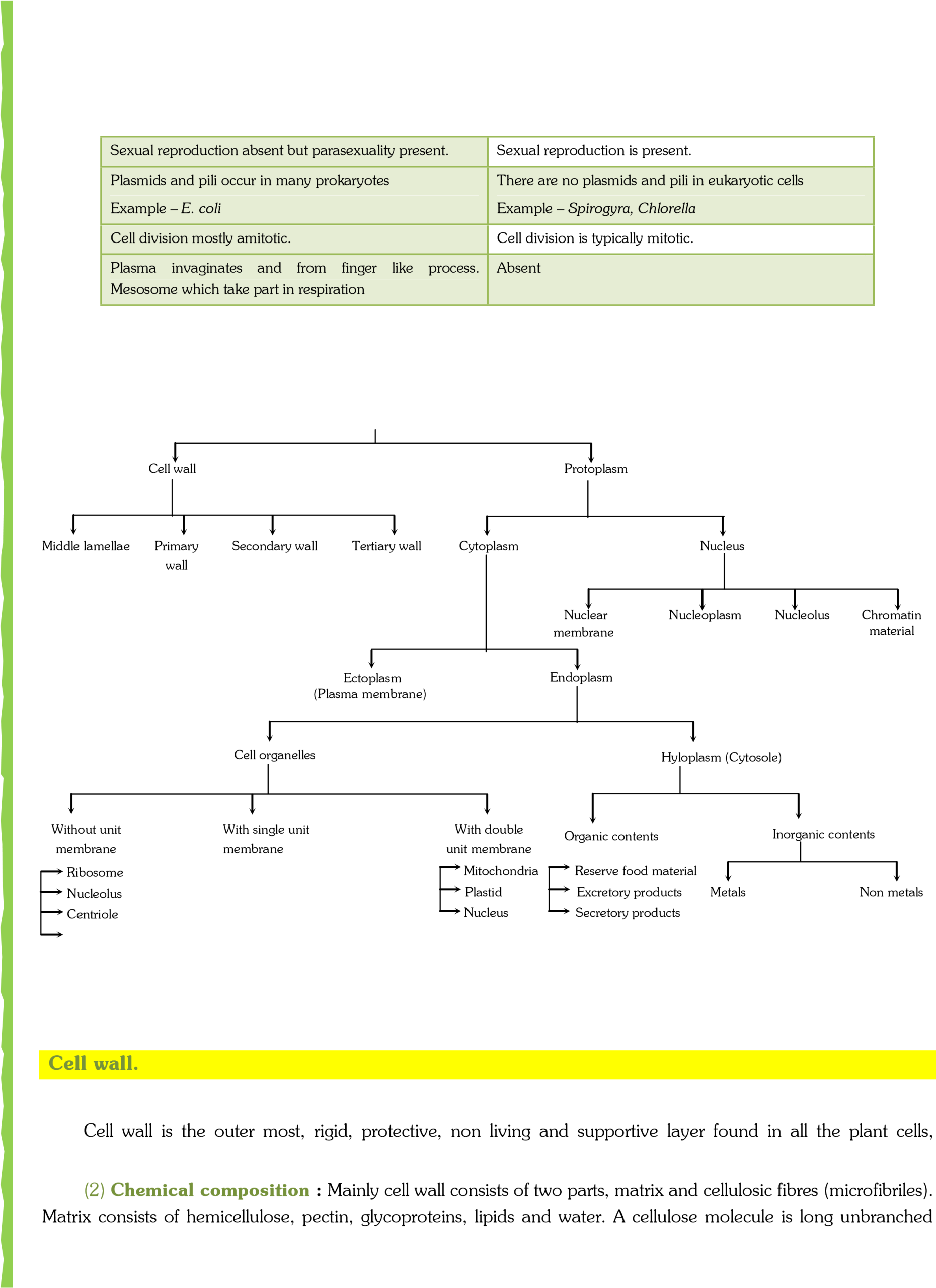
(3) Structure : Cell wall consists of middle lamella, primary wall, secondary wall, tertiary wall.
| middle lamella. Pectin is used as commercial jellying agent. Which is present outside the primary wall. | L.S. cell walls of two adjacent cells
Fig : Layers of cell wall in T.S. and L.S. of a cell |
- Middle lamella : Middle lamella is the outermost region which functions as a cementing layer between
Middle Lamella
Primary Wall
Lumen
T. S. of A Plant cell
Middle Lamella
Primary Wall
S
1
S
1
S
2
S
2
S
3
S
3
two cells. It is absent on the outer free Secondary wall Layers surface. It ruptures to create intercellular spaces. Middle lamella is
formed of calcium and magnecium Secondary wall
Layers pectate. Fruit softening is due to gelatinisation of pectic compounds of
- Primary wall : A young plant cell forms a single layer of wall material. This layer is known as the primary cell wall. The primary wall is thin, elastic and capable of expansion in a growing cell. It grows by intussusception. Meristematic and parenchymatous cells have primary cell wall only. The cells of leaves and fruits too have only primary wall.
- Secondary wall : In mature cell, more layers of wall material are added internal to the primary wall. These are called the secondary cell wall. Growth by addition of new wall material on the primary wall is called accretion. The secondary wall is thick and rigid. It usually consists of three layers, which are often named S1,S2 and S .3 It is found in collenchyma and sclerenchyma cells, xylem vesseles.
- Tertiary wall : Sometimes tertiary wall is laid down on secondary wall, e.g., tracheids of gymnosperms. It is composed of cellulose and xylan, another ploysaccharides.
(4) Origin : A cell wall is organised at telophase stage of cell division. The plane and place of cell wall is determined by the microtubules. Fragments of ER and vesicles of golgi body alligned at the equator, called as phragmoplast, later which forms the cell plate. The synthesis of cellulose takes place by the help of enzyme cellulose synthase present in the plasma membrane.
The cell plate forms the cell wall. A cell posses three phases of growth namely cell formation, cell elongation and cell maturation. The formation of new cells occurs by mitotic activity. The cell elongation is initiated by an increase in cell turgor. It is brought about by special proteins called expansion. They are of two types α−expansion and β−expansion. As a result, lacunae or gaps appear in between the cellulose micelle. There are two possibilities for the deposition of new wall material.
- By intussuception : As the cell wall stretches in one or more directions, new cell wall material secreted by protoplasm gets embedded within the original wall.
- By apposition : In this method new cell wall material secreted by protoplasm is deposited by definite thin plates one after the other.
Differences between primary and secondary cell wall
| Primary cell wall | Secondary cell wall | ||
| Primary wall is laid inner to middle lamella | Secondary wall is laid inner to primary wall. | ||
| It is formed in a growing cell. | It is formed when the cells have stopped growing. | ||
| It is capable of extension. | Extensibility is absent except in collenchyma cells. | ||
| It is single layered. | It is three or more layered. | ||
| Cellulose content is comparatively low (5 – 20%). | Cellulose content is comparatively high (20 – 90%). | ||
| Cellulose microfibrils are shorter, wavy and loosely arranged. | They are longer, closely arranged straight and parallel. | ||
| Protein content up to 5%. | Protein content up to 1%. | ||
| Hemicellulose content is high up to 50%. | It is 25% of the total. | ||
| Lipid content up to 5 – 10%. | Lipid is absent. | ||
Primary wall is comparatively thin 1 – 5 |
It is comparatively thick 5 – 10 |
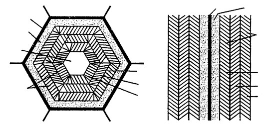
(5) Thickenings of cell wall : In many secondary walls specially those of xylem the cell wall becomes hard and thick due to the deposition of lignin. With the increasing amount of lignin, deposition protoplasm is lost. First the lignin is deposited in middle lamella and primary wall and later on in secondary wall. Like cellulose lignin is permeable to water and substances dissolved in it. Lignin is deposited at specific places of the cell walls due to which xylem tracheids and trachea take up following forms: Fig : Different types of secondary wall thickenings –
A
B
C
D
E
F
- Annular thickenings : Deposition of lignin takes (a) annular (b) spiral (c) scalariform (d) reticulate (e) pittedplace in the form of rings on the inner surface of protoxylem simple pits (f) pitted-bordered pit
cells. These rings are placed one above the other leaving some space in between each other.
- Spiral thickenings : In these thickenings deposition of lignin takes place in the form of complete spiral bands and are formed in tracheids and trachea of protoxylem.
- Scalariform (Ladder like) thickenings : In these thickenings lignin is deposited in the form of transverse rods of the ladder. The unthickened areas between the successive thickenings appear as elongated transverse pits. This type of thickening is common in protoxylem.
- Reticulate (Net like) thickenings : The lignin is deposited in the form of a net or reticulum. The unthickened areas are irregular in shape. These are found in metaxylem.
- Pitted thickenings : These are found in metaxylem. In such thickening the whole inner wall is more or less uniformly thickened leaving here and there some unthickened areas called pits.
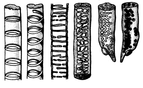
(6) Pits : Secondary walls may have irregular thickenings at some places and these places are called pits. Pits are of two types :–
- Simple pit : In which pit chamber is uniform in diameter.
- Bordered pit : In which pit chamber is flask shaped in tracheids of gymnosperm and vessels of angiosperms.
Bordered pit
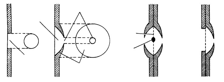
Simple pit
Pit chamber
Pit cavity
Pit aperture
Border
Margo
Torus
A. Simple pit B. Bordered pit C. Bordered pit pair D. Half bordered pit
- Plasmodesmata : Tangle (1879) first of all discovered them and were studied elaborately by Strasburger (1901). A number of plasmodesmata or cytoplasmic strands are present in pit through which the cytoplasm of one cell is in contact with another. Endoplasmic reticulum plays a role in origin of plasmodesmata.
- Intercellular spaces : In mature cells certain spaces or cavities are produced which are of 3 types.
- Schizogenous cavities : In mature cells, the cell walls separate from each other and form a cavity. e.g., resin canals in Pinus.
- Lysogenous cavities : It is formed by the break down of cell walls e.g., Citrus oil cavities.
- Schizo-lysogenous cavities : Both the above processes are involved in this cavity formtion e.g., protoxylem of maize.
(9) Function of cell wall : The cell wall serves many functions –
- It maintain shape of the cells.
- It protect the cells from mechanical injury.
- It wards off the attacks of pathogens (viruses, bacteria, fungi, protozoans).
- It provides mechanical support against gravity. It is due to the rigid cell walls that the aerial parts of the plants are able to keep erect and expose their leaves to sunlight.
- The cell wall prevents undue expansion of the cell when water enters by osmosis to compensate for the lack of contractile vacuole. This prevents bursting of cells.
- It allows the materials to pass in and out of the cell.
- Though permeable, the cell wall plays some regulatory role on the passage of materials into and out of the cell.
- Many enzymic activities associated with metabolism are known to occur in the cell wall.
- Cutin and suberin deposits check loss of water form the cell surface by evaporation.
- The cell wall helps in the maintenance of balance of intracellular osmotic pressure with that of its surroundings.
- Pores in the cell walls permit plasmodesmata to link up all the protoplasts into a system called symplast (symplasm).
- The walls of xylem vessels, tracheids and sieve tubes allow movement of materials.
- The wall in some cases has a role in defence and offence by means of spines.
- Growth of the cell wall enables the cells to enlarge in size.
- Cell wall and intercellular spaces constitute a nonliving component of plant body known as apoplasm.
Important Tips
- Peptidoglycane = murein = mucopeptide is the only cell wall material of prokaryotes. It’s sugar portion consists of NAG and NAM.
- In fungi cell wall is made up of chitin (polymer of N- acetyl glucosamine). In bacteria it is composed of protein lipid polysaccharide having N-acetyl glucosamine (NAG) and N-acetyl muramic acid (NAM). • Cell wall proteins –
HRGP – Hydroxy proline rich glycoprotein → Phloem and cambium.
PRP– Proline rich protein → Xylem, fibres, cortex.
GRP– Glycine rich protein → Xylem.
| Plasma membrane. |
- Definition : Every living cell is externally covered by a thin transparent electron microscopic, elastic regenerative and selective permeable membrane called plasma membrane. It is quasi fluid in nature. According to Singer and Nicolson it is “protein iceberg in a sea of lipid”. A cell wall lies external to plasmalemma in plant cells, many monerans, some protists and fungal cells. Membranes also occur inside the cells. They are collectively called biomembranes. The term cell membrane was given by C. Nageli and C. Cramer (1855) for outer membrane covering of the portoplast. It was replaced by the term plasmalemma or plasma membrane by Plowe (1931).
- Chemical composition : Proteins lipoprotein (Lipid +Protein) are the major component forming 60% of the plasma membrane. Proteins provide mechanical strength and responsible for transportation of different substances. Proteins also act as enzyme. Lipids account may 28%-79% depending upon the type of cell and organism involved (in humans, myelin 79%). Because of the presence of lipids, membranes are always continuous, unbroken structures and are deformable and their over all shape can change. The lipids of plasma membrane are of three types namely phospholipids, glycolipids and sterols. A glycolipid may be cerebroside or ganglioside. The sterol found in the membrane may be cholesterol (Animals), phytosterol (Plants) or ergosterol (Microorganisms). A lipid molecule is distinguishable into a head of glycerol and two tails of fatty acids.
Carbohydrates form 2%–10%. Oligosaccharides are the main carbohydrates present in plasma membrane. The carbohydrates of plasma membrane are covalently linked to both lipid and protein components. The common sugars found in the plasma membrane are D – glucose, D – mannose, D – glactose, N – acetyl glucosamine, N – acetyl galoactosamine (Both are amino sugars) and sialic acid. Generally the terminal sugar of oligosaccharides is sialic acids (Also known as N – acetylneuraminic acid NANA) which gives them a negative charge.
- Ultra structure : Under electron microscope the plasma membrane appears three layered, i.e. trilaminar or tripertite. One optically light layer is of lipid and on both sides two optically dense protein layers are present.
Generally the plasma membrane is 75 Å thick (75 – 100Å), light layer is 35 Å while dark layers are 20 Å 20+ Å in thickness.
- Molecular structure and different models : Several models have been proposed to explain the structure and function of the plasma membrane.
- Overton’s model : It suggests that the plasma membrane is composed of a thin lipid bilayer.
- Sandwich model : It was proposed by Davson and Danielli (1935). According to this model the light biomolecular lipid layer is sandwiched between two dense protein layers. This model was also said to be unit membrane hypothesis.
- Robertson’s unit membrane model : It states that all cytoplasmic membranes have a similar structure of three layers with and electron transparent phospholipid bilayer being sandwiched between two electron dense layer of proteins. All biomembranes are either made of a unit membrane or a multiple of unit membrane. Its thickness is about 75 Å with a central lipid layer of 35 Å thick and two peripheral protein layers of 20 Å thick.
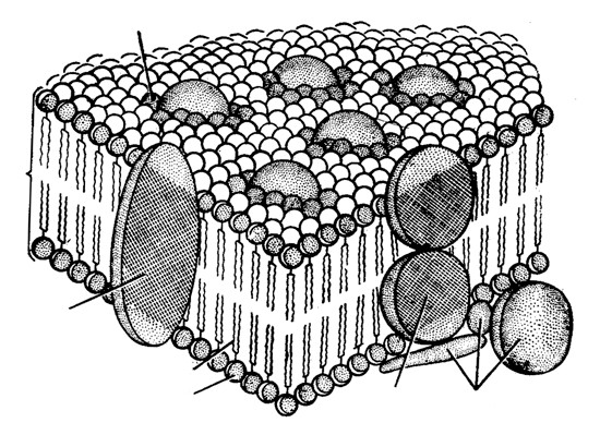
- Fluid mosaic model : The most important and widely accepted latest model for plasma membrane was given by Singer and Nicolson in 1972. According to them it is “protein iceberg in a sea of lipids.”
According to this model, the cell membrane consists of a highly viscous fluid matrix of two layers of phospholipid molecules. These serve as relatively impermeable barrier to the passage of most water soluble molecules. Protein molecules occur in the membrane, but not in continuous layer; Instead, these occur as separate particles asymmetrical arranged in a mosaic pattern.
Boundary lipid
Intrinsic protein
Hydrophobic tail
Hydrophilic head
Intrinsic
protein
Extrinsic
proteins
Lipid
bilayer
Lipid
Polar end
Non-polar end
Fig : Fluid
–
mosaic model of the plasma membrane. Proteins floating in a
Some of these are loosely bound at the polar surfaces of lipid layers, called peripheral or extrinsic proteins. Others penetrate deeply into the lipid layer called integral or intrinsic proteins. Some of the integral proteins penetrate through the phospholipid layers and project on both the surface. These are called trans membrane or tunnel proteins (glycophorins). Singly or in groups, they function as channels for sea of lipid. Some proteins span the lipid bilayer, others are exposed
passage of water ions and other solutes. only to one surface or the other (Modified after De Robertis et al.; 1975). The channels may have gate mechanism for opening in response to specific condition. The carbohydrates occur only at the outer surface of the membrane. Their molecules are covalently linked to the polar heads of some lipid molecules (forming glycolipids) and most of the proteins exposed at outer surface (forming glycoproteins).
The sugar protions of glycolipids and glycoproteins are involved in recognition mechanisms :–
- Sugar recognition sites of two neighbouring cells may bind each other causing cell to cell adhesion. This enables cells to orientate themselves and to form tissues.
- Through glycoproteins, bacteria recognise each other. e.g., female bacteria are recognised by male bacteria.
- These provide the basis of immune response and various control system, where glycoproteins act as antigens. Lipid and integral proteins are amphipathic in nature i.e., they have hydrophilic and hydrophobic groups with in the same molecules. The NMR (Nuclear magnetic resonance) and ESR (Electron spin resonance) studies showed that the membrane is dynamic. The lipid tails show flexibility. The molecule can rotate or show flip flop motion.
Difference between protein types
| Extrinsic Protein | Intrinsic Protein |
| These are associated with surface only. | These lie throughout phospholipid matrix and project on both surfaces, also called transmembrane or tunnel protein. |
| They form about 30% of the total membrane protein. | They form about 70% of total membrane proteins. |
| Example – Spectrin in red blood cells & ATPase in mitochondria. | Example – Rhodopsin in retinal rod cells. |
(5) Membrane protein can be of following types with different functions
- Carrier molecules : These bind with the specific molecules into or out of the cell. This provides selective exchange of materials. The carrier protein molecules are called “permeases” e.g., Na+ – K+ pump, Na+– sugar transport.
- Receptor molecules : The glycoproteins on the cell surface act as receptors that recognize and bind with specific molecules.
- Enzyme molecules : The inner mitochondrial membrane carrier enzyme comprising the electron transport chain for cellular respiration.
(6) Cell membranes are fluid and dynamic due to (i) The constituent molecules can move freely in the membrane.
- The cell membranes are constantly renewed during the cells life.
- They can repair minor injuries.
- They expand and contract during cell movement and during change in shape.
- They allow interactions of cells such as recognition of self and fusion of cells.
(7) Membrane permeability : According to permeability, membranes are classified as –
- Permeable membrane : They allow both solvent and solute molecules or ions through them. e.g., cellulose wall, lignified cell walls.
- Impermeable membrane : They do not allow solute and solvent molecules. e.g., heavily cutinised or suberinised cell walls in plants.
- Semi-permeable membrane : They allow solvent molecules only. e.g., membranes of colloidion, parchment paper and copper ferrocyanide membranes.
- Differentially permeable membrane : All membranes found in plants allow some solutes to pass through them along with the solvent molecules. e.g., plasma membrane, tonoplast (vacuolar membrane) etc.
(8) Intercellular communications/modification of plasma membrane/following structures are derived from plasma membrane
- Microvilli : They are fingers like evaginations of 0.1 µmdiameter, engaged in absorption. e.g., intestinal cells, hepatic cell, mesothelial cells. The surface having microvilli is called striated border or brush border.
- Lomasomes : They are plasmalemma foldings found in fungal cells.
- Mesosomes : It serves as site for cellular respiration in prokaryotes.
- Tight junctions : Plasma membrane of two adjacent cells are fused at a series of points with a network of ridges or sealing strands. e.g., capillaries, brain cells collecting tubules etc.
- Plasmodesmata : They are protoplasmic bridges amongst plant cells, which occur in area of cell wall pits. It was discovered and reported by Tangle and Strasburger respectively.
- Desmosomes : concerned with cell adherence.
(9) Functions
- They control the flow of material through them and provides passage for different substances.
- It is differentially permeable, solute particles (1-15 Å) can pass through it.
- It is not only provides mechenical strength but also acts as a protective layer.
- Plasma membrane is responsible for the transportation of materials, molecules, ions etc.
- It helps in osmoregulation.
- Diffusion of gases take place through plasma membrane by simple and facilitated diffusion.
- Transport of ions, small polar molecules through active (energy used) and passive transport (energy not used).
- Gases like O2and CO2 diffuse rapidly in solutions through membranes.
- Ions and small polar molecules diffuse slowly through the membranes.
- Some solute molecules or ions first bind with certain specific carrier or transport proteins called permeases.
- Water as well as some solute molecules and ion pass through membranes pores; pores are always bordered by channel proteins.
- When diffusion takes place through channel, called simple diffusion and through carrier proteins, called facilitated diffusion.
(10) Membrane transport : It is passage of metabolites, by-products and biochemicals across biomembrane. Membrane transport occurs through four methods–passive, facilitated, active and bulk. Size of the particles passing through plasmalemma is generally 1 – 15 Å.
(i) Passive transport : No energy spent. Passive transport occurs through diffusion and osmosis.
- Diffusion : It is movement of particles from the region of their higher concentration or electrochemical potential to the region of their lower concentration or electrochemical potential. Electrochemical potential operates in case of charged particles like ions. Diffusion can be observed by opening a bottle of scent or ammonia in one corner, placing a crystal of copper sulphate in a beaker of water or a crystal of KMnO4 on a piece of gelatin. Simple diffusion does not require carrier molecules.
Independent Diffusion : In a system having two or more diffusion substances, each individual substance will diffuse independent of others as per gradient of its own concentration, diffusion pressure or partial pressure form region of higher one to region of lower one.
Rate of diffusion is proportional to difference in concentration and inversely to distance between the two ends of the system, inversely to square root of relative density of substance and density of medium, directly to temperature and pressure.
- Osmosis is diffusion of water across a semipermeable membrane that occurs under the influence of an osmotically active solution.
- Mechanism of passive transport : Passive transport can continue to occur if the absorbed solute is immobilised. Cations have a tendency to passively pass from electropositive to electronegative side. While anions can pass from electronegative to electropositive side. There are two modes of passive transports.
Lipid matrix permeability : Lipid soluble substances pass through the cell membrane according to their solubility and concentration gradient, e.g., triethyl citrate, ethyl alcohol, methane.
Hydrophillic membrane channels : They are narrow channels formed in the membrane by tunnel proteins. The channels make the membrane semipermeable. Water passes inwardly or outwardly from a cell through these channels according to osmotic gradients. CO2and O2also diffuse through these channels as per their concentration gradients. Certain small ions and other small water soluble solutes may also do so.
- Ultrafiltration is fine filtration that occurs under pressure as from blood capillaries, epithelia and endothelia. It is of two types : –
- Paracellular through leaky junctions or gaps in between cells.
- Transcellular through fenestrations in the cells. ‘Dialysis’ is removal of waste products and toxins from blood by means of diffusion between blood and an isotonic dialysing solution.
(e) Facilitated transport or Facilitated diffusion : It is passage of substances along the concentration gradient without expenditure of energy that occurs with the help of special permeating substances called permeases. Permeases form pathways for movement of certain substances without involving any expenditure of energy. At times certain substances are transported alongwith the ones requiring active transport. The latter phenomenon called cotransport. Facilitated transport occurs in case of some sugars, amino acids and nucleotides.
(ii) Active transport : It occurs with the help of energy, usually against concentration gradient. For this, cell membranes possess carriers and gated channels.
- Carrier particles or Proteins : They are integral protein particles which have affinity for specific solutes. A solute particles combines with a carrier to form carrier solute complex. The latter undergoes conformational change in such a way as to transport the solute to the inner side where it is released into cytoplasm.
- Gated channels : The channels are opened by either change in electrical potential or specific substances, e.g., Calcium channels.
Active transport systems are also called pumps, e.g., H+pump, K+pump, Cl− pump, Na+ −K+ pump. The pumps operate with the help of ATP. K+ −H+ exchange pump occurs in guard cells. Na+ −K+ exchange pump operates across many animal membranes. For every ATP hydrolysed, three Na+ ions are passed out while two K+ions are pumped in. Sea Gulls and Penguins operate Na+ −K+ pump for excreting NaCl through their nasal glands.
Active transport of one substance is often accompanied by permeation of other substances. The phenomenon is called secondary active transport. It is of two main types, cotransport (e.g., glucose and some amino acids alongwith inward pushing of excess Na+ ) and counter-transport (Ca2+ and H+movement outwardly as excess Na+ passes inwardly).
(iii) Bulk transport : It is transport of large quantities of micromolecules, macromolecules and food particles through the membrane. It is accompanied by formation of transport or carrier vesicles. The latter are endocytotic and perform bulk transport inwardly. The phenomenon is called endocytosis. Endocytosis is of two types, pinocytosis and phagocytosis. Exocytic vesicle perform bulk transport outwardly. It is called exocytosis. Exocytosis performs secretion, excretion and ephagy.
- Pinocytosis : (Lewis, 1931). It is bulk intake of fluid, ions and molecules through development of small endocytotic vesicles of 100 – 200 nm in diameter. ATP, Ca2+,fibrillar protein clathrin and contractile protein actin are required. Fluid-phase pinocytosis is also called cell drinking. It is generally nonselective. For ions and molecules the membrane has special receptor or adsorptive sites located in small pits. They perform adsorptive pinocytosis. After coming in contact with specific substance, the area of plasma membrane having adsorptive sites, invaginates and forms vesicle. The vesicle separates. It is called pinosome. Pinosome may burst in cytosol, come in contact with tonoplast and pass its contents into vacuole, form digestive vacuole with lysosome or deliver its contents to Golgi apparatus when it is called receptosome.
- Phagocytosis : (Metchnikoff, 1883). It is cell eating or ingestion of large particles by living cells, e.g., white blood corpuscles (neutrophils, monocytes), Kupffer’s cells of liver, reticular cells of spleen, histiocytes of connective tissues, macrophages, Amoeba and some other protists, feeding cells of sponges and coelentrates. Plasma membrane has receptors. As soon as the food particle comes in contact with the receptor site, the edges of the latter evaginate, form a vesicle which pinches off as phagosome.
One or more lysosomes fuse with a phagosome, form digestive vacuole or food vacuole. Digestion occurs inside the vacuole. The digested substances diffuse out, while the residual vacuole passes out, comes in contact with plasma membrane for throwing out its contents through exocytosis or ephagy.
Important tips
- E. Grater and H. Grendel (1926) : Proposed leaflet model which states that plasma membrane is formed of bilayer sheet of phospholipids.
- Wolpers (1941) : Proposed lattice model which states lipids are distributed in a framework of proteins.
- Hilleir and Hoffman (1953) : Proposed micellar model. Plasma membrane is formed of micelles of lipid molecules.
- Sandwich model of Danielli and Davson (1935) is based on physical and chemical properties.
- Proteins of plasma membrane provide functional specificity, elasticity and mechanical support.
- The arrangement of phospholipid molecules in bilayer forms a water resistant barrier. • Glycoproteins of plasma membrane determine antigen specificity of cell. These glycoproteins from major histocompatible complex (MHC) which are of specific type in every individual so act as finger print of the cell.
- Negative charge of the membrane is due to N – acetyl neuraminic acid (NANA)/sialic acid.
- Lehninger described the percentage of extrinsic and intrinsic protein.
- Harmone receptor proteins of plasma membrane of target cells act as signal transduction.
- Phospholipids show asymmetric distribution in plasma membrane lacithin and sphingomycelin mainly found in outer phospholipids layer while cephalin and phosphatidyl serine are mainly present in inner phosphalipid layer.
- Lomasomes : Infolds of plasma membrane found in fungi. These were reported by Moore and Mclean.
- Transosomes found in follicular cells of ovary of birds and have triple unit membrane. First reported by Press(1964).
- Lipid soluble substances pass through the plasma membrane move readily than the water soluble substances.
- Term biomembrane was coined by Singer and Nicolson.
- Nehar and Sakmann discovered ion-channels in plasma membrane and they were awarded Noble prize for it in 1971.
- Pinocytosis and phagocytosis do not take place in prokaryotic cells.
- Singer and Nicolson’s model differs from Robertson’s model in the arrangement of proteins.
- Plasma membrane contains ATPase enzymes.
- Plasma gel or ectoplasm are the synonyms of plasma membrane.
- The secondary structure of the integral protein buried in the lipid bilayer of a cell membrane is nature.
| Protoplasm. |
(1) Definition : Protoplasm is a complex, granular, elastic, viscous and colourless substance. It is selectively or differentially permeable. It is considered as “Polyphasic colloidal system”. (2) Discoveries
-
- J. Huxley defined it as “physical basis of life”.
- Dujardin (1835) discovered it and called them “sarcode”.
- Purkinje (1837) renamed it as “Protoplasm”.
- Hugo Von Mohl (1844) gave the significance of it.
- Max Schultz (1861) gave the protoplasmic theory for plants.
- Fischer (1894) and Hardy (1899) showed its colloidal nature.
- Altman (1893) suggested protoplasm as granular.
- Composition : Chemically it is composed of
| Water | 75 – 85% | Carbon | 20% |
| Proteins | 10 – 25% | Oxygen | 62% |
| Lipids | 2 – 3% | Hydrogen | 10% |
| Inorganic Materials | 1% | Nitrogen | 3% |
Trace elements – 5% (Ca, ,P Cl, ,S K, Na,Mg, ,I Fe, etc.)
Maximum water content in protoplasm is found in hydrophytes, i.e. 95% where as minimum in seeds, spores (dormant organs) i.e. 10 – 15%. In animals water is less (about 65%) and proteins are more (about 15%).
-
- Physical properties of protoplasm : Cyclosis movement are shown by protoplasm. These are of two types.
- Rotation : In one direction, either clockwise or anticlockwise e.g., Hydrilla, Vallisneria. Found only in eukaryotes.
- Circulation : Multidirectional movements around vacuole e.g. Tradescantia.
- It shows stimulation or irritability.
- Protoplasm is polyphasic. Colloidal substance or true solution because true solution act as dispersion medium and different colloidal particles constitute dispersed phase.
- It shows increased surface area and adsorption.
- It shows sol – gel transformation.
- It is highly viscous.
- It coagulates at 60o C or above or if treated with concentrated acids or bases.
- It shows Brownian movements.
- It’s specific gravity is slightly more than 1.
- It’s pH is on acidic side, but different vital activities occur at neutral pH which is considered as 7, injury decreases the pH of the cell (i.e. 5.2 – 5.5) and if it remains for a long time, the cell dies.
- Scattering and dispersion of light is shown by protoplasm i.e. Tyndall effect.
| Cytoplasm. |
The substance occur around the nucleus and inside the plasma membrane containing various organelles and inclusions is called cytoplasm.
-
- The cytoplasm is a semisolid, jelly – like material. It consists of an aqueous, structureless ground substance called cytoplasmic matrix or hyaloplasm or cytosol.
- It forms about half of the cell’s volume and about 90% of it is water.
- It contains ions, biomolecules, such as sugar, amino acid, nucleotide, tRNA, enzyme, vitamins, etc.
- The cytosol also contains storage products such as glycogen/starch, fats and proteins in colloidal state.
- It also forms crystallo – colloidal system.
- Cytomatrix is differentiated into ectoplasm or plasmagel and endoplasm or plasmasol.
- Cytomatrix is three dimensional structure appear like a network of fine threads and these threads are called microfilaments (now called actin filaments or microtrabecular lattice) and it is believed to be a part of cytoskeleton. It also contains microtubules and inter mediate cytoplasmic filaments.
- Hyaloplasm contains metabolically inactive products or cell inclusions called deutoplast or metaplasts.
- Cytoplasmic organelles are plastid, lysosome, sphaerosome, peroxisome, glyoxysomes, mitochondria, ribosome, centrosome, flagellum or cilia etc.
- The movement of cytoplasm is termed as cyclosis (absent in plant cells).
| Mitochondria. |
(1) Definition : (Gk – mito = thread ; chondrion = granule) Mitochondria are semi autonomous having hollow sac like structures present in all eukaryotes except mature RBCs of mammals and sieve tubes of phloem. These are absent in all prokaryotes like bacteria and cyanobacteria. Mitochondria are also called chondriosome, chondrioplast, plasmosomes, plastosomes and plastochondriane.
(2) Discoveries
- These were first observed in striated muscles (Voluntary) of insects as granules by Kolliker (1850), he called them “sarcosomes”.
- Flemming (1882) called them “fila” for thread like structure.
- Altman (1890) called them “bioplast”.
- C. Benda (1897) gave the term mitochondria.
- F. Meves (1904) observed mitochondria in plant (Nymphaea).
- Michaelis (1898) demonstrated that mitochondria play a significant role in respiration.
- Bensley and Hoerr (1934) isolated mitochondria from liver cells.
- Seekevitz called them “Power house of the cell”.
- Nass and Afzelius (1965) observed first DNA in mitochondria.
- Number of mitochondria : Presence of mitochondria depends upon the metabolic activity of the cell.
Higher is the metabolic activity, higher is the number e.g., in germinating seeds.
-
- Minimum number of mitochondria is one in Microasterias, Trypanosoma, Chlorella, Chlamydomonas (green alga) and Micromonas. Maximum numbers are found (up to 50,000) in giant Amoeba called Chaos – Chaos. These are 25 in human sperm, 300 – 400 in kidney cells and 1000 – 1600 in liver cells.
- Mitochondria of a cell are collectively called chondriome.
- Size of mitochondria : Average size is
0.5–1.00 µm and length up to 1 – 10 µ m.
-
- Smallest sized mitochondria in yeast cells (1µm3 ).
- Largest sized are found in oocytes of Rana pipiens and are 20 – 40 µm.
- A dye for staining mitochondria is Janus B – green.
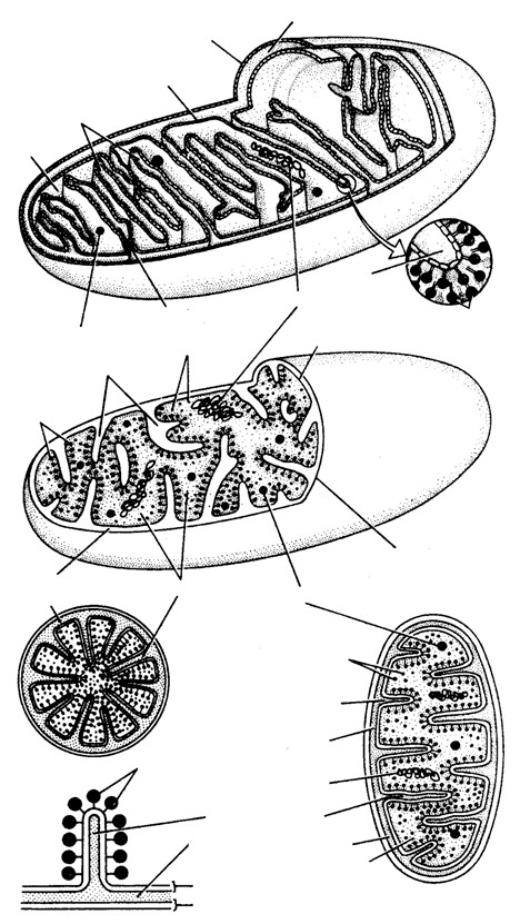
- Ultrastructure of mitochondria : Mitochondrion is bounded by two unit membranes separated by perimitochondrial space (60 – 80 Å). The outer membrane is specially permeable because of presence of integral proteins called porins. The inner membrane is selective permeable. The inner membrane is folded or convoluted to form mitochondrial crests. In animals these are called cristae and in plants these folding are called tubuli or microvili.
The matrix facing face is called ‘M’ face and face towards perimitochondrial space is called ‘C’ face. The ‘M’ face have some small stalked particles called oxysomes or F1 particle or elementory particle or Fernandez – Moran Particles. Each particle is made up of base, stalk and head and is about 10nm in length. Number of oxysomes varies to 104 to 105 per mitochondrion and chemically they are made of
1
Intermembranous space
Outer membrane
Inner membrane
Cristae
Matrix
Inclusions
Intercristaeal space
Tubuli
Ribosomes
F
Particles or
Oxysomes
DNA
Inner membrane
Crista
A
F
1
Particles or
Oxysomes
Intermembranous
space
B
Matrix
Inclusions
Outer membrane
Matrix
Ribosomes
Outer
chamber
DNA
Intratubuli space
Intermembranous
space
Inner
chember
Inner membrane
Outer membrane
C
D
F
1
Particles or
Oxysomes
Fig : Three dimentional structure of mitochondrion. A. From an animal cell. B. From plant cell, C. T.S.
mitechondrion, D. One tubule
Perimitochondrial space
F
1
Particles
Outer membrane
Inner membrane
Mitochondrial crest
Respiratory chain
and enzymess
Outer chamber
Protein layer
Lipid layer
Intracristael space
F
1
Particles
Fig : Molecular organization of inner membrane of mitochondria
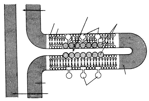
phospholipid core and protein cortex. Oxysomes have ATPase enzyme molecule (Packer, 1967) and therefore, responsible for ATP synthesis. These elementary particles are also called F0 – F1 particles.
In the matrix 2–6 copies of naked, double stranded DNA (circular) and ribosome of 70 S type are present. It is rich in G-C ratio. Basic histone proteins are absent in mitochondrial DNA. The synthesis of ATP in mitochondria is called oxidative phosphorylation, which is O2 dependent and light independent. Cristae control dark respiration. F0 particles synthesize all the enzymes required to operate Kreb’s cycle. Inner membrane contains cytochrome.
(6) Semi-autonomous nature of mitochondrion : Mitochondria contain all requirements of protein synthesis :
- 70 S ribosomes.
- DNA molecules to form mRNA and also replicate.
- ATP molecules to provide energy.
The mitochondria can form some of the required proteins but for most of proteins, these are dependent upon nuclear DNA and cytoplasmic ribosomes, so the mitochondria are called semi-autonomous organelles.
- Two states of mitochondria : When ATP synthesis is low or the respiratory chain of mitochondrion is inhibited, it is called inactive or orthodox state, and has large amount of matrix and only a few cristae. But when mitochondria are active or condensed state, and have small amount of matrix and highly developed cristae. This shows that the development of mitochondria depends upon the physiological activity of the cell.
- Chemical composition : Cohn gave the chemical composition of mitochondrion:
Proteins = 65 – 70%
Lipids = 25 – 30% (90% phospholipids and 10% cholesterol, Vit. E., etc)
2 – 5% RNA Some amount of DNA
The mitochondrial matrix has many catabolic enzymes like cytochrome oxidase and reductases, fatty acid oxidase, transaminase, etc.
(9) Enzymes of Mitochondria
- Outer membrane : Monoamine oxidase, glycerophosphatase, acyltransferase, phospholipase A.
- Inner membrane : Cytochrome b,c1,c,a, (cyt.b, cyt.c1, cyt.c, cyt.a, cyt.a3) NADH, dehydrogenase, succinate dehydrogenase, ubiquinone, flavoprotein, ATPase.
- Perimitochondrial space : Adenylate kinase, nucleoside diphosphokinase.
- Inner matrix : Pyruvate dehydrogenase, citrate synthase, aconitase, isocitrate dehydrogenase, fumarase, α−Ketoglutarate dehydrogenase, malate dehydrogenase.
(10) Origin : Mitochondria are self-duplicating organelles due to presence of DNA molecules so new mitochondria are always formed by growth and division of pre-existing mitochondria by binary fission.
Difference between outer and inner membrane of mitrochondria
| Outer membrane | Inner membrane |
| It is smooth having less area. | It is infolded to form cristae hence large surface area. |
| It is freely permeable. | Semipermeable, impermeable to coenzyme A and NAD. |
| It consist 50% lipid and 50% protein. | It consist 80% protein and 20% lipid. |
| Sialic acid is more (4 – 5 time). | Sialic acid is less. |
| Near about 14% enzymes are present. | Near about 60 enzymes are present. |
(11) Functions of mitochondria
- Mitochondria are called power house or storage batteries or ATP mills as these are sites of ATP formation.
- Intermediate products of cell respiration are used in the formation of steroids, cytochromes, chlorophyll, etc.
- These are also seat of some amino acid biosynthesis.
- Mitochondria also regulate the calcium ion concentration inside the cell.
- Site of Krebs cycle and electron transport system.
- Site of thermiogenesis.
- Yolk nucleus (a mitochondrial cloud and golgi bodies) controls vitellogenesis.
- Mitochondria of spermatid form nebenkern (middle piece) of sperm during spermiogenesis.
- It is capable of producing its own DNA.
- Mitochondria release energy during respiration.
- Mitochondria contain electron transport system.
Important Tips
- Petite character in yeast and cytoplasmic male sterility in maize are examples of mitochondrial inheritance.
- Mitochondria are believed to be bacterial endosymbionts.
- Mitochondria show a large degree of autonomy or independence in their functioning.
- Mitochondria as a place of cellular respiration were first observed by Hogeboom. Enzymes of Kreb’s cycle or TCA cycle or citric acid cycle are present in matrix except succinic dehydrogenase which is found attached to inner mitochondrial membrane.
- With the help of phase contrast microscope mitochondria has been studied well.
- Mitochondria can be separated by centrifugation.
- Mitochondria are called as “cell inside cell” by Schiff (1982).
- Life of mitochondria is not more than 5 days.
- Mitochondria are yellowish due to riboflavin.
- 70% of total enzymes of a cell are found in mitochondria.
- Mitochondrial genome has 200 kilobase pairs.
- Mitochondria has the similarity , with bacteria as both have 70 S ribosome, circular DNA and RNA.
- Mitochondria are rich in manganese.
- It has its own electron transport system.
- Mitochondria and chloroplasts have many resemblances.
- According to endosymbiotic origin of mitochondria by Kirns Altman, mitochondria were intially a free living, aerobic bacteria which during to the process of evolution entered an anaerobic cell and become established as mitochondria. This theory is supported by many similarities which exist between bacteria and mitochondria.
- Lehninger discovered oxysomes.
- Percentage of mitochondrial DNA in cells is 1% of the total cellular DNA.
- Parson discovered stalkless and hollow spherical particles present on outer surface of outer mitochondrial membrane. • When mitochondria treated with detergents like digitonin or lubral, their outer unit membrane is removed and remaining part is called Mitoplast
- The F1 particle is made up of five types of subunits namely αβγδ, , , and ε. of these αis heaviest andεis lightest.
- In prokaryotic cell, plasma membrane infolding makes a structure mesosome. Which is analogous structure of mitochondria of eukaryotic cell (both part in respiration).
| Plastids. |
(1) Definition : Plastids are semiautonomous organelles having DNA, RNA, Ribosomes and double membrane envelope which store or synthesize various types of organic compounds as ATP and NADPH + H+ etc. These are largest cell organelles in plant cell.
(2) History
- Haeckel (1865) discovered plastid, but the term was first time used by Schimper (1883).
- A well organised system of grana and stroma in plastid of normal barley plant was reported by de Von Wettstein.
- Park and Biggins (1964) gave the concept of quantasomes.
- The term chlorophyll was given by Pelletier and Caventou, and structural details were given by Willstatter and Stall.
- The term thylakoid was given by Menke (1962).
- Fine structure was given by Mayer.
(3) Types of plastids : According to Schimper, Plastids are of 3 types: Leucoplasts, Chromoplasts and Chloroplasts.
Leucoplasts : They are colourless plastids which generally occur near the nucleus in nongreen cells and possess internal lamellae. Grana and photosynthetic pigments are absent. They mainly store food materials and occur in the cells not exposed to sunlight e.g., seeds underground stems, roots, tubers, rhizomes etc. These are of three types.
- Amyloplast : Synthesize and store starch grains. e.g., potato tubers, wheat and rice grains.
- Elaioplast (Lipidoplast, Oleoplast) : They store lipids and oils e.g. castor endosperm, tube rose, etc.
- Aleuroplast (Proteinoplast) : Store proteins e.g., aleurone cells of maize grains.
Chromoplasts : Coloured plastids other than green are kown as chromoplasts. These are present in petals and fruits, imparting different colours (red, yellow, orange etc) for attracting insects and animals. These also carry on photosynthesis.
These may arise from the chloroplasts due to replacement of chlorophyll by other pigments e.g. tomato and chillies or from leucoplasts by the development of pigments.
All colours (except green) are produced by flavins, flavenoids and cyanin. Cyanin pigment is of two types one is anthocyanin (blue) and another is erythrocyanin (red). Anthocyanin express different colours on different pH value. These are variously coloured e.g. in flowers. They give colour to petals and help in pollination. They are water soluble. They are found in cell sap.
Green tomatoes and chillies turn red on ripening because of replacement of chlorophyll molecule in chloroplasts by the red pigment lycopene in tomato and capsanthin in chillies. Thus, chloroplasts are changed into chromatophores.
Chloroplast : Discovered by Sachs and named by Schimper. They are greenish plastids which possess photosynthetic pigments.
- Number : It is variable. Number of chloroplast is 1 in Spirogyra indica, 2 in Zygnema, 16 in S.rectospora, up to 100 in mesophyll cells. The minimum number of one chloroplast per cell is found in Ulothrix and species of Chlamydomonas.
- Shape : They have various shapes
| Shape | Example |
| Cup shaped | Chlamydomonas sp. |
| Stellate shaped | Zygnema. |
| Collar or girdle shaped | Ulothrix |
| Spiral or ribbon shaped | Spirogyra |
| Reticulate | Oedogonium |
| Discoid | Voucheria |
- Size : It ranges from 3 – 10 µm (average 5 µm) in diameter. The discoid chloroplast of higher plants are 4 – 10µm in length and 2– 4µm in breadth. Chloroplast of spirogyra may reach a length of 1 mm. Sciophytes
(Shade plant) have larger chloroplast.
- Chemical composition :
- Proteins 50 – 60%,
- Lipids 25 – 30% ,
- Chlorophyll – 5- 10 %,
- Carotenoids (carotenes and xanthophylls) 1 –2%,
- DNA – 0.5%, RNA 2 – 3%,
- Vitamins K and E,
- Quinines, Mg, Fe, Co, Mn, P, etc. in traces.
(v) Ultrastructure : It is double membrane structure. Both membranes are smooth. The inner membrane is less permeable than outer but rich in proteins especially carrier proteins. Each membrane is 90 – 100 Å thick. The inter-membrane space is called the periplastidial space. Inner to membranes, matrix is present, which is divided into two parts.
(a) Grana : Inner plastidial membrane of the chloroplast is invaginated to form a series of parallel membranous sheets, called lamellae, which form a
number of oval – shaped closed sacs, called thylakoids. Frets or Lamellae
Thylakoids are structural and functional elements of Outer chloroplasts. These thylakoids contain all the membrane requirements of light reactions e.g., pigments like chlorophyll, carotenoids, plastoquinone, plastocyanin, Inner
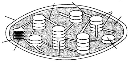
Granum in L.S.
Thylakoid
Stroma
Granum
membrane etc. that are involved in photosynthesis. Each thylakoid has an intrathylakoid space, called loculus (size 10-30Å)
Fig : A chloroplast in section (diagrammatic) bounded by a unit membrane. Along the inner side of thylakoid membrane, there are number of small rounded para-crystalline bodies, called quantasomes (a quantasome is the photosynthetic unit) which can trap a mole of quantum of light and can bring about photosynthetic act. Each quantasome contains about 230 chlorophyll molecules and 50 carotenoid molecules.
In eukaryotic plant cells, a number of thylakoids are superimposed like a pile of coins to form a granum. The number of thylakoids in a granum ranges from 10-100 (average number is 20-50). The number of grana per chloroplast also varies widely e.g., one granum per chloroplast in Euglena while there are 40-60 grana per chloroplast in spinach. The size of each granum varies from 0.2 – 0.6µm in diameter. But in blue-green algae, the thylakoids are not organised to form granum.
Adjacent grana are interconnected by branched tubules, called stromal lamellae or Fret-channel or Fret membrane’s.
(b) Stroma : It is transparent, proteinaceous and watery substance. Dark reaction of photosynthesis occurs in this portion. Stroma is almost filled with “Rubisco” (about 15% of total enzyme, protein) enzyme CO2is accepted by this enzyme. CO2 assimilation results in carbohydrate formation. It has 20 – 60 copies of naked circular double stranded DNA. Each DNA copy is 40µ in length, which can code for 125 amino acids. All plastids of a cell called as
“Plastidome” (Dangeared 1920) in stroma. Amount of DNA per chloroplast is 10–15 g. Chloroplast genome has 145 kilobase pairs. It shows semiautonomous nature and ribosomes are of 70 S type.
(vi) Pigments of chloroplast : Willsttater and Stall observed the following pigments:
- Chlorophyll a : C55 H72 O N Mg5 4 (with methyl group)
- Chlorophyll b : C55 H70 6 4O N Mg (with aldehyde group)
- Chlorophyll c : C35 32 5H O N Mg4
- Chlorophyll d : C54 H70 O N Mg6 4
- Carotenes, Xanthophylls : Carotenoids.
Difference between Chl. a and Chl. b
| Chl. a | Chl. b |
| Absorption peak at 430, 662. | It is 453, 642. |
| Bluish green in colour. | Yellowish green. |
| Soluble in petroleum, ether. | Soluble in methyl alcohol. |
| Functional group at C3 position is CH3 | Functional group attached to pyrrol ring is CHO. |
| Present in all green plants excepts autotrophic bacteria. | Present in all green plants except blue green, brown and red algae. |
| In chloroplast it is 75%. | It is 25% |
| In reflected light Chl. a shows blood red colour while in transmitted light, it shows blue green colour. | In reflected light it show dull brown colour while in transmitted light, it shows yellowish green colour. |
(vii) Chlorophylls and their presence : Term by Cavantou (1818). It’s molecule has tetrapyrollic or porphyrin head (15 Å ×15 Å ) and phytol tail (20 Å long). Mg++ is present in the centre of porphyrin head. If chlorophyll is burnt only Mg is left.
- Chlorophyll b : It is found in members of chlorophyceae.
- Chlorophyll c : It is found in members of phaeophyceae, bacillariophyceae.
- Chlorophyll d : It is found in members of rhodophyceae.
- Chlorophyll e : It is found in members of xanthophyceae.
- Phycoerythrin and phycocyanin (phycobilins) are the red and blue green pigments in rhodophyceae and cyanophyceae respectively.
- Fucoxanthin (brown pigment) in phaeophyceae.
- Bacteriochlorophyll (C55 74 6 4H O N Mg) or chlorobium chlorophyll present in photosynthetic bacteria.
These pigment are red in acidic and blue in alkaline medium.
(viii) Carotenoids : These are hydrocarbons, soluble in organic solvents. These are of 2 types:
- Carotenes : C40 56H derivatives of vitamin A. Carrot coloured αβγ, , carotene, lycopene, etc.
- Xanthophyll : C40 56 2H O ,yellowish in colour, fucoxanthin, violaxanthin. Molar ratio of carotene and xanthophyll in young leaves is 2 : 1.
(ix) Plastids are interchangeable
Leucoplast Chloroplast
Chromoplast
(degenerate chloroplast)
The leucoplast and chloroplast are interconvertible but once they have converted into chromoplast, the reverse can not take place. Because, chromoplasts are aged or degenerated form of chloroplast e.g. in tomato.
| Young ovary (colourless) | – | Leucoplast |
| Young fruit (green) | – | Chloroplast |
| Mature fruits (red) (due to Lycopene) | – | Chromoplast. |
| In carrot leucoplast | – | Chromoplast (carotene) etc. |
(x) Origin of chloroplast : Plastids, like the mitochondria, are self duplicating organelles. These develop from colourless precursors, called proplastids. They are believed to be evolved from endosymbiont origination.
(4) Function of plastids
- It is the site of photosynthesis, (light and dark reaction).
- Photolysis of water, reduction of NADP to NADPH2 take place in granum.
- Photophosphorylation through cytochrome b6 f, plastocyanine and plastoguinone etc.
- They store starch or factory of synthesis of sugars.
- Chloroplast store fat in the form of plastoglobuli.
- They can be changed into chromoplasts to provide colour to many flowers and fruits for attracting animals.
- They maintain the percentage of CO2 and O2 in atmosphere.
Important Tips
- Murphy and Leech (1978) have reported the synthesis of fatty acids in the spinach chloroplast.
- Proplastids are precursor of all type of plastids.
- Capasanthin is the pigment in carotenoids found in bacteria, fungi and chilly.
- Solar energy is trapped in lamella by chlorophylls but in bacteria trapping centre is B890.
- The chloroplast with nitrogen fixing genes (nif genes) constitute nitroplast.
- Pyrenoids : A proteinaceous core around which starch is deposited mostly found in the chloroplast of algae and in some bryophytes.
- Algal classification is based on pigmentation pattern.
- Eye spot or stigma is photosensitive carotenoid pigment.
- Intact chloroplast can be separated by sugar solution (2.5 M).
- Mitochondria and plastids both have own DNA molecules which is called as Extranuclear/ Extrachromosomal DNA.
- Plastids are absent from monerans, fungi and animals these are also absent from gametes and zoospores of plants.
- Ris and Plaut (1962) reported DNA in chloroplast and was called plastidome. It forms about 0.5% of total cellular DNA and is rich in G-C pairs.
- Plastidoribosomes : Ribosomes of plastids and are of 70S type. These were reported by Jacobson et. al. (1963)
- Thylakoid term was given by Menke (1961).
- Transducers : Structure which are involved in energy transformations e.g. mitochondria and plastids.
- Plastids are the largest cell organelles. The plastids in the order of their increasing size are Chloroplast→Chromoplast→Elaioplast→Aleuroplast→Amyloplast
- Quantasome is formed of 160 chlorophyll a + 70 chlorophyll b molecules and 50 carotenoid molecules.
- Scattered thylakoids in the cytoplasm of cyanobacteria and photosynthesis bacteria are known as chromatophores.
- Chromatophore term was given by Schmitz.
| Endoplasmic reticulum (ER). |
- Definition : It is well developed electron microscopic network of interconnected cisternae, tubules and vesicles present throughout the cytoplasm, especially in the endoplasm.
- Discovery : Garnier (1897) was first to observe the ergastoplasm in a cell. The ER was first noted by Porter, Claude, and Fullman in 1945 as a network. It was named by Porter in 1953.
- Occurrence : The ER is present in almost all eukaryotic cells. A few cells such as ova, embryonic cells, and mature RBCs, however, lack ER. It is also absent in prokaryotic cell.
In muscle cells, it is called sarcoplasmic reticulum, myeloid bodies and nissel granules are believed to be formed from ER. ER is little develop in meristematic cells.
- Chemical composition : All the components of ER are lipoperoteins and trilaminar like the plasma membrane but differ in following
- Thinner (50 − 60 Å) than plasma membrane.
- With less cholesterol.
- With more lipids.
- The lumen is filled with fluid containing 70% phospholipids lecithin and cephalin etc.
(5) Ultrastructure : The ER is made up of three components :
- Cisternae : These are flattened, unbranched, sac like structures. They lie in stacks (piles) parallel to one another. They bear ribosomes. They contain glycoproteins named ribophorin-I and ribophorin-II that bind the ribosomes. Found in protein forming cells.
- Vesicles : These are oval or rounded, vacuole like elements, scattered in cytoplasm. These are also studded with ribosomes.
- Tubules : Wider, tubular, branched elements mainly present near the cell membrane. They are free from ribosomes. These are more in lipid forming cells.
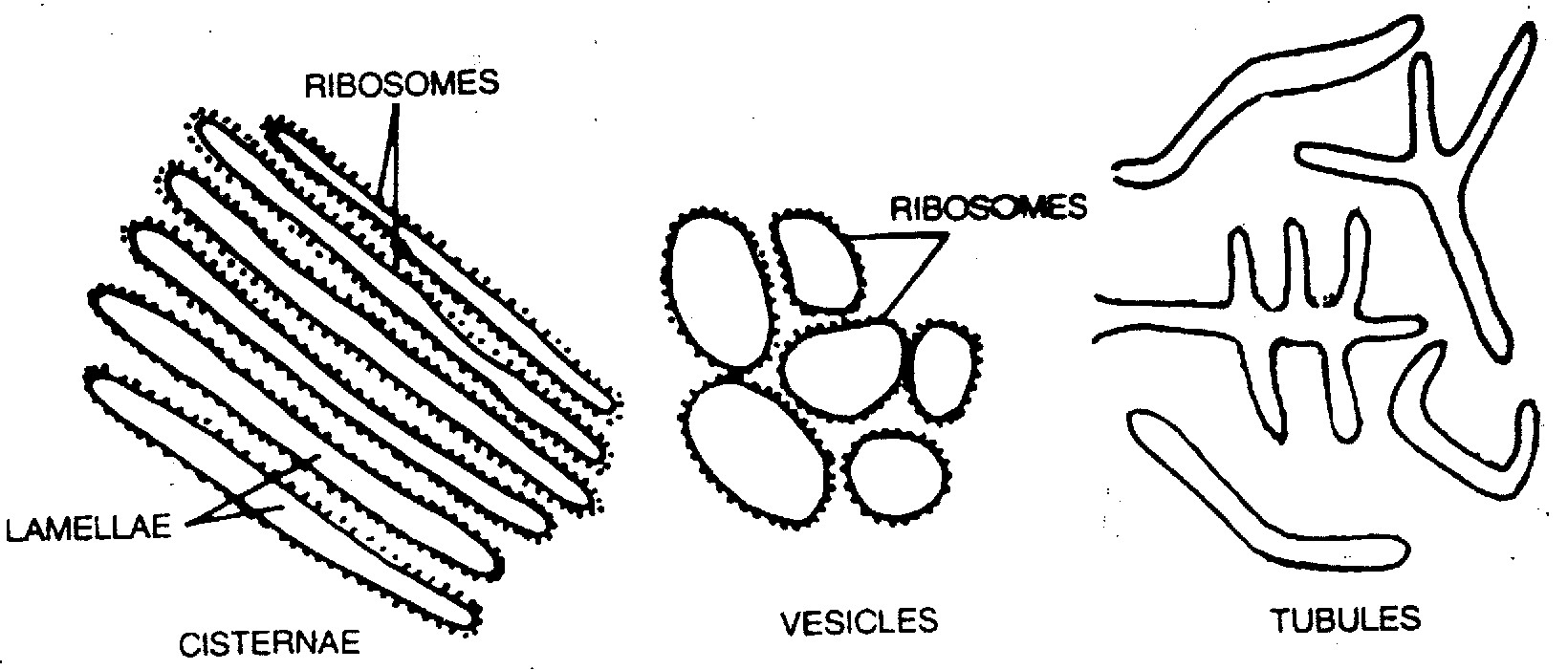
Ribosomes
Ribosomes
Vesicles
Lamellae
Cisternae
Tubules
Fig : Elements of Endoplasmic Reticulum
All the three structures are bound by a single unit membrane.
(6) Types of ER : Depending upon the presence of ribosomes, the ER has been categorised into two types:
- A smooth or Agranular endoplasmic reticulum (SER) : It consists mainly of tubules and vesicles. It has no ribosomes associated to it. It is well developed in the muscle cells, adipose tissue cells, interstitial cells, glycogen storing liver cells, etc. and the cells that synthesize and secrete steroids. SER also takes part in synthesis of vitamins, carbohydrates and detoxification. Detoxification of pollutants carcinogens and drugs is carried out SER of liver cells and mitochondria, SER is associated with storage and release of Ca2+ions. It gives rise to spherosomes.
- Rough or Granular endoplasmic reticulum (RER) : It mainly consists of cisternae. It has ribosomes attached on its cytoplasmic surface. It is abundant in cells engaged in production and excertion of proteins, e.g., plasma cells, goblets cells, pancreatic acinus cells and certain liver cells. The RER is more stable than SER. The RER is basophilic due to the presence of ribosomes. Ribosomes are attached to ER through hydrophobic interaction.
The proteins synthesised by the ER membrane bound ribosomes pass into the ER lumen, where most of the proteins are glycosylated. For this, an oligosaccharide is always linked to the − NH2group on side chain of an asparagine residue. The ER lumen serves as a compartment to contain substances which must be kept separate from cytosol. In the ER lumen, the enzymes modify the proteins.
Differences between SER and RER
| SER | RER |
| SER or smooth endoplasmic reticulum does not possesses ribosomes over the surface of its membrane. | RER possesses ribosomes attached to its membrane. |
| It is mainly formed of vesicles and tubules. | It is mainly formed of cisternae and few tubules. |
| It is engaged in the synthesis of glycogen lipids and steroids. | The reticulum takes part in the synthesis of proteins. |
| Pores are absent so that materials synthesised by SER do not pass into its channels. | RER possesses narrow pores below its ribosomes for the passage of synthesised polypeptides into ER channels. |
| SER is often peripheral. It may be connected with plasmalemma. | It is often internal and connected with nuclear envelope. |
| Ribophorins are absent. | RER contains Ribophorins I and II for providing attachment to ribosomes. |
| SER gives rise to sphaerosomes. | It helps in the formation of lysosome through the agency of golgi apparatus. |
(7) Origin : RER is formed from nuclear membrane while SER is formed from RER by loss of ribosomes. Rough vesicles originate only from RER after homogenisation of cell. RER breaks in small fragments (Vesicles) and it is called microsome (This is not a cell organelle). ER constitute cytoskeleton and also help as intracellular transport system. And it is sensitive to irritation. (8) Functions
- Synthesis and secretion of specific proteins via – golgi bodies.
- Formation of protein ribophorin. Which helps in attachment of ribosome.
- Give rise to SER.
- Provides surface for synthesis of cholesterol, steroid, ascorbic acid and visual pigments.
- It helps in synthesis of harmones e.g., testosterone and estrogen.
- It helps in glycogenolysis in the liver cells and brings about detoxification (SER).
- Gastric cells secreting zymogen have well developed SER.
- ER is a component of cytoskeleton (Spread as a net) of cell and provides mechanical support and shape to the cell.
- ER acts as segregation apparatus and divides the cytoplasm into chambers. Compartmentalisation is most necessary for cellular life.
- It participates in the formation of cell-plate during cytokinesis in the plant cells by the formation of phragmoplasts.
- ER has many types of enzymes e.g. ATPase, reductases, dehydrogenases and phosphatases.
(9) Sacroplasmic reticulum : It is a modified SER striated muscle fibres which forms a network of interconnected tubules in the sarcoplasm. It helps in conduction of motor nerve impulses throughout the muscle fibre and in the removal of lactic acid so prevents muscle fatigue. It is called “ergastoplasm” in muscle and “nisslegranules” in nerve cells.
Important Tips
- Annullated lamellae : It was first reported by Mc Culloch (1952) in the egg of sea urchin. Formed by blebbing of outer nuclear membrane.
- Transitional ER : It is RER without ribosomes.
- Microsome : This term was used by Claude (1941). It probably refers to these fragments of ER which are associated to ribosomes.
- Sjostrand gave the term α−cytomembrane for RER.
- Veratti (1902) reported sacroplasmic recticulum in the muscle fibers.
- Nissl’s granules are the masses of RER in the cyton of neurons.
- Myeloid bodies are the masses of tubules (S0 SER) found in retinal cells and are related with photoreception.
- Total ER in the cell – 2/3 RER + 1/3 SER.
- In rapidly dividing cells endoplasmic reticulum is poorly developed.
| Golgi complex. |
- Definition : Golgi complex is made up of various membranous system e.g. cisternae, vesicles and vacuoles. These are also called golgi bodies, golgisomes, lipochondrion, dictyosomes, Dalton complex, idiosomes or Baker’s body. These are also called “traffic police” of the cell.
- Discovery : First observed by George (1867) but it’s morphological details were given by Camillo Golgi (1898), in nerve cells of barn fowl and cat.
 Occurence : It is present in all eukaryotic cells. They form 2% of total cell volume. In a cell these are found above centriole or near nucleus. In plants, these are scattered irregularly in the cytoplasm and called as “dictyosomes”. These are absent in bacteria and blue green algae, RBCs, spermatozoa of bryophytes and pteridophytes, and sieve tube cells of phloem of angiosperm.
- Size and number : The size of the golgi body varies with the metabolic state of cell and hence it is called pleomorphic. Large in mature functional and secretary cell e.g., germinal cells, goblet cells, but small size in non-secretary cells. There may be
25,000 dictysomes present in rhizoidal cells of Chara. Fig : Arrangement of membrane, tubles and vesicles in golgi complex Average number 10 – 20 per cell. Number increases during cell division.
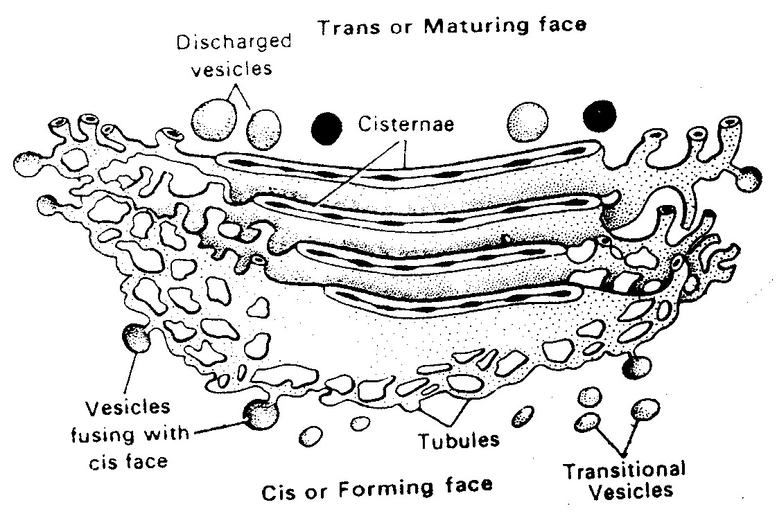
- Structure : Under transmission electron microscope the st. of golgibodies was study by Dalton and Felix (1954), golgi body is made of 4 parts.
- Cisternae : Golgi apparatus is made up of stack of flat. Sac like structure called cisternae. The margins of each cisterna are gently curved so that the entire golgi body takes on a cup like appearance. The golgi body has a definite polarity. The cisternae at the convex end of the dictyosome comprises forming face (F. face) or cis face. While the cisternae at the concave end comprises the maturing face (M. face) or trans face. The forming face is located next to either the nucleus or endoplasmic reticulum. The maturing face is usually directed towards the plasma membranes. It is the functional unit of golgi body.
- Tubules : These arise due to fenestration of cisternae and it forms a complex of network.
- Secretory vesicles : These are small sized components each about 40 Å in diameter presents along convex surface of edges of cisternae. These are smooth and coated type of vesicles. Smooth or secretory vesicles, which have a smooth surface and contain secretions of the cell and coated vesicles, that have rough surface. They carry materials to or from the cisternae.
- Golgian vacuoles : These are spherical components each about 600 Å in diameter. These are produced by vesiculation of saccules of cisternae. Scattered cisternae are called dictyosomes and condition is called diffused.
(6) Function
- The main function of golgi body is secretion, so it is large sized among the secretory cells. Secretion are released either by exocytosis or reverse pinocytosis.
- Glycosidation of lipids i.e. addition of oligosaccharides to produce glycolipids.
- Glycosylation of proteins i.e. addition of carbohydrate to produce glycoproteins.
- Formation of lysosomes.
- Golgi body forms the cell plate. During cell division by secreting hemicellulose formation of enzyme and hormones (Thyroxine) etc.
- Matrix of connective tissue is formed by golgi complex.
- In oocytes of animal, golgi apparatus functions as the centre around which yolk is deposited i.e. vitellogenesis.
- Membrane of the vesicles produced by golgi apparatus join in the region of cytokinesis to produce new plasmalemma.
- It is also called export house of cell.
- Golgi body contains phospholipids, proteins, enzymes and vitamin-c.
- The golgi complex gives rise to the acrosome in an animal sperm.
(7) Origin : Most accepted view is that golgi body originates from RER-that has lost its ribosomes from this RER arise transport vesicles that contain Golgi membrane and fuse with the saccule on the forming face of Golgi apparatus. This is why this face is called the forming face. Important Tips
- According to Camillo Golgi “Apparato reticulare interno” (internal reticular apparatus) is Golgi body.
- Cellulose, hemicellulose and pectin are synthesized by Golgi body.
- Metal silver impregnation technique was used by Camillo Golgi.
- Sperm acrosome is made of golgi apparatus.
- The main enzyme of golgi complex are glycosyl transferase, nucleoside diphosphatase and thiamine pyrophosphatase.
- Zymogen is processed in it.
- Term “trophospongium” given by Holmgen.
- The number of golgi bodies increase during cell division. Phragmoplast is the precursor of cell plate.
- The basophilic ergastoplasm in gland cells indicate the richness of golgi bodies.
- Root cap cells are rich in golgi complex secreting mucilage, which lubricates the root tip.
- Proteins and fats are stored in vacuoles and vesicles of golgi complex.
- In fungi, unicisternal dictyosomes are found.
- Zone of exclusion : A zone of clear cytoplasm with no ribosomes, mitochondria etc. around the golgi body.
- Perner gave the term dictyosome.
- Mollenhaver and Whaley (1963): Reported polarity in golgi complex.
- GERL : Golgi-endoplasmic reticulum-lysosome system.
- GER : Golgi associated endoplasmic reticulum.
| Lysosomes. |
- Definition : Lysosomes are electron microscopic, vesicular structures of the cytoplasm, bounded by a single membrane which are involved in intracellular digestive activities, contains hydrolytic enzymes, so called lysosomes.
- Discovery : These were first discovered by a Belgian biochemist, Christian de Duve (1995) in the liver cells and were earlier named pericanalicular dense bodies. Terms Lysosome was given by Novikoff under the study of electron microscope. Maltile (1964) was first to demonstrate their presence in plants, particularly in the fungus neurospora.
- Occurrence : These are absent from the prokaryotes but are present in all eukaryotic animal cells except mammalian RBCs. They have been recorded in fungi, euglena, cotton and pea seeds.
- Shape : These are generally spherical in shape but are irregular in plant root tip cells.
- Size : Size range is 0.2-0.8 µm while size is 0.5 µm (500 nm).
- Number : Lysosomes are more in those cells which are involved in intracellular digestive activities e.g., WBCs of blood, histiocytes of connective tissue; phagocytes of liver and spleen; osteoclasts; cells of degenerating tissue like tail of tadpole larva etc.
- Ultrastructure : Under electron microscope, a lysosome is formed of two parts :
- Limiting membrane : It is outer, single layered, lipoproteinous and trilaminar unit membrane. It keeps a limit on glycoproteinous digestive enzymes.
- Matrix : It is inner, finely granular and highly heterogeneous group substance inside the membrane.
(8) Types : The lysosomes change the nature of their contents at different times in the same cell. This variation is referred to as polymorphism. On the basis of their contents, four types of lysosomes are recognised.
- Primary Lysosomes : A newly formed lysosome contains enzymes only. It is called the primary lysosomes. Its enzymes are probably in an inactive state.
- Secondary Lysosomes : When some material to be digested enters a primary lysosome, the latter is named the secondary lysosome, or phagolysosome or digestive vacuole, or heterophagosome. This commonly occurs by fusion of a primary lysosome with a vacuole (pinosome or phagosome) or a secretory granule.
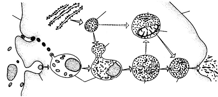
Plasma
membrane
Primary lysosome
or storage granule
Endoplasmic
reticulum
Plasma
membrane
Autophagic
vacuole
Digested
mitochondrion
Secondary
lysosome
Phagosome
Digestive
vacuole
Residual
body
Defecation or
exocytosis or wastes
Food particles taken
in by endocytosis
Fig : Different types of lysosomes and their origin
- Tertiary lysosomes/Residual bodies : In a secondary lysosome, the enzymes digest the incoming materials. The products of digestion pass through the lysosome membrane into the cytoplasmic matrix for use as a source of nutrition or energy. Indigestible matter remains in the secondary lysosome. A secondary lysosome containing indigestible matter is known as the residual body or tertiary lysosome. The latter meets the cell by exocytosis (ephagy).
- Autophagosomes /Autolysosomes /Autophagic vaculoes : A cell may digest its own organelles, such as mitochondria, ER. This process is called autophagy or autolysis. These are formed of primary lysosomes. The enzymes (hydrolytic) of lysosomes digest the organelles thus enclosed. Therefore, the lysosome are sometimes called disposal units/suicidal bags.
(9) Chemical composition : Matrix of primary lysosome is formed of hydrolases, which is involved in hydrolysis or polymeric compounds, that operate in acidic medium at pH 5, so called acid hydrolases. Upto now 50 types of enzyme have been reported to be present in latent form in different types of lysosomes. These enzymes are synthesized on RER, transported to cisternae of golgi body where these are packed into the lysosomes. These are as (i) Proteases e.g., cathepsin and collagenase.
- Nucleases e.g., DNAse and RNAse.
- Glycosidases e.g., β-galactosidase, β-glucoronidase.
- Phosphatases e.g., ATPase, acid phosphatase (marker enzyme).
- Sulphatases e.g., for sulphate-linked organic compounds.
- Esterases e.g., phospholipase, acid lipase.
(10) Origin : Lysosomes arise from the golgi complex their membrane and hydrolytic enzymes are synthesized on the RER and are transported invesicles to the golgi complex for modification and packaging.
(11) Functions
- Lysosomes take part in digestion of food through phagosomes, known as intracellular digestion.
- In metamorphosis of many animals certain embryonic parts are digested by it.
- Obstructing structures are destroyed by lysosome.
- Lysosomes perform the function of exocytosis and endoytosis.
- Lysosomes of sperms provide enzyme for breaking limiting membrane of egg e.g., hyaluronidase enzyme.
- They cause breakdown of ageing and dead cells.
- Lysosomes functions as trigger of cell division or initiate cell division by digesting repressor molecules.
- Nucleases (DNAse) of lysosomes may cause gene mutations which may cause disease like leukemia or blood cancer (partial deletion of 21st chromosome).
- Sometimes residual bodies accumulate inside the cells leading to storage diseases e.g. a glycogen storage disease called Pompe’s disease, polynephritis Hurler’s disease (deformed bones due to accumulation of mucopolysaccharides).
- Lysosomes also engulf the carcinogens.
Important Tips
- Cholesterol, cortisol and cortisone acts as a stablizers of lysosomal membrane, while absence of oxygen, X-rays UV rays and excess of vitamin A and E act as labilizers and weaken the lysosomal membrane.
- Polymorphism in lysosomes were described by De Robertis et. al (1971).
- Lysosomes can hydrolyse all types of organic compounds except cellulose.
| Ribosomes. |
-
- Definition : The ribosomes are smallest known electron microscopic without membrane, ribonucleo–protein particles attached either on RER or floating freely in the cytoplasm and are the sites of protein synthesis.
- Discovery : In 1943 Claude observed some basophilic bodies and named them as microsome. Palade
(1955) coined the term ribosome (form animal cell). Ribosomes in nucleoplasm were observed by Tsao and Sato (1959). First isolated by Tissieres and Watson (1958) from E. coli. Ribosomes found in groups are termed as polyribosomes or ergosomes (Rich and Warner 1963 observed first time polyribosomes).
- Occurrence : These are found in both prokaryotes as well as eukaryotes these are present only in free form in the cytoplasm. While in the eukaryotes the ribosomes are found in two forms in the cytoplasm, free form and bind form (bound on RER and outer nuclear membrane). These are also reported inside some cell organelles like mitochondria and plastids respectively called mitoribosomes and plastidoribosomes.
- Number : The number of ribosomes depends upon the RNA contents of the cell. These are more in plasma cells, liver cells, Nissl’s granules of nerve cells, meristematic cells and cancerous cells.
- Types of ribosomes : It is determined on the basis of sedimentation coefficient measured in Svedberg unit or ‘S’ unit and their size. Velocity of sedimentation is 1×10⁻¹³ cm / sec/ dyne/gm.
- 70S ribosomes : Found in prokaryotes, mitochondria and plastid of eukaryotes. Each is about 200 – 290Å × 170 – 210Å in size and 2.7 ×10⁶ dalton in molecular weight.
- 80S ribosomes : Found in cytoplasm of eukaryotes. Each is about 300 – 340 Å × 200 – 240 Å in size and 4.5 – 5.0 ×10⁶ daltons in molecular weight.
- 77S, 60S and 55S ribosomes : Levine and Goodenough (1874) observed 77S ribosomes in fungal mitochondria 60S ribosomes in animal mitochondria and 55S in mammalian mitochondria.
- Structure : Each ribosome is formed of two unequal subunits, which join only at the time of protein synthesis. In 70S and 80S ribosomes, 50S and 30S, 60S and 40S are larger and smaller subunits respectively. Larger subunits is dome shaped and attached to ER by glycoproteins called “ribophorins”. It has a depression on the flate side which leads into a channel having elongating polypeptide chain. It has a protuberance, a ridge and a stalk. It also has 2 binding sites. Peptidyl or P or Donor site and Amino actyl or A or Acceptor site.
30S Subunit 40S Subunit
300
–
340
Å Length
200-240
Å Width
290
Å Length
50S Subunit 60S Subunit
210Å Width
70S Ribosome 80S Ribosome
Fig : 70S and 80S ribosome
These sites are for the attachment of charged tRNA molecules. Smaller subunit is oval shaped and fits as a cap on flat side of larger subunit. It has a platform, cleft head and base. It has binding site for mRNA. Delimiting membrane is not found in it. Ribosomes are attached to ER through hydrophobic interactions.
- Chemical composition : Ribosomes are chemically composed of rRNA and proteins Ribonucleo-Protein (RNP). Lipids are altogether absent in ribosomes. Ribosomes are strongly negative binding cations and basic dyes. 70S ribosomes has 60-65% rRNA and 35-40% proteins (ratio is 1.5 : 1). rRNAs are of three types : 23S type and
5S type rRNAs in 50S and 16S type rRNA in 30S sub-units. There are about 55 types of proteins in 70S ribosome out of which 21 proteins are found in 30S while 34 proteins are found in 50S ribosomal sub-unit and are called core-proteins.
80S ribosome has 45% rRNA and 55% proteins (ratio is about 1 : 1). r-RNA are of four types : 28S, 5S and 5.8S types of rRNAs in 60S and 18S type rRNA in 40S sub-units. There are about 70 types of proteins in 80S ribosome out of which 30 proteins are found in 40S while 40 proteins are found in 60S ribosomal sub-units. The ribosomal proteins are basic and almost surround the rRNA. Some proteins act as structural proteins while other proteins act as enzymes e.g., peptidyl transferase of 50S (controls the interlinking of amino acids by peptide bonds).
A 1 10× −3 (0.001 M) molar concentration of Mg++ is needed for the structural cohesion of ribosomes i.e., for holding the two subunits together. If this concentration is increased by ten folds, two ribosomes unite to form a dimer. The sedimentation coefficient of dimer of 70S ribosmes is 100S and that of 80S is 120S. By decreasing the
Mg++ conc. to normal, the dimer breaks into monomers (single ribosomes).
+Mg++ +Mg++
70 70S+ S ++ 100DimerS, 80S+ 80S –Mg++ 120S Monomers –Mg
If the Mg++ concentration is decreased to 1 10× −4 molar, the ribosomes break up into its sub-units. The 70S ribosome breaks up into 50S and 30S sub-units. These 50S and 30S sub-units further dissociates into RNA and protein components. Similarly, the 80S ribosomes dissociates into 60S and 40S sub-units which further breakup into RNA and protein components.
- Biogenesis of ribosome :
- In eukaryotes the ribosomal RNAs like 18S, 5.8S and 28S are synthesized by nucleolus and 5S RNA out of the nucleus.
- In prokaryotes both rRNA and its protein are synthesized as well as assembled by cytoplasm.
- Polyribosomes or Polysomes : When many ribosomes (generally 6 – 8) are attached at some mRNA strand. It is called polysome. The distance between adjacent ribosomes is of 90 nucleotides. These are functional unit of protein synthesis.
- Function :
- Ribosomes are also called protein factory of the cell or work branch of proteins.
- Free ribosomes synthesize structural proteins and bounded ribosomes synthesize proteins for transport.
- Ribosomes are essential for protein synthesis.
- Help in the process of photosynthesis.
- They are found numerously in actively synthesizing cells like liver cells, pancreas, endocrine, yeast cells and meristematic cells.
- Ribosomes also store the proteins temporarily.
- These also store rRNAs, which helps in protein synthesis.
- Enzyme peptidyl transferase occurs in large subunit of ribosome which helps in protein synthesis.
- Newly formed polypeptide is protected from degradation by cytoplasmic enzymes in large sub-unit of ribosomes before releasing it into RER lumen. Important tips
- Gunter Blobel and David Sabatini of Rockfeller university proposed signal hypothesis in 1971. Both scientist has been awarded the Nobel prize (1999) for this protein signalling.
- Ultra-structure of ribosomal subunits was given by James A. Lake (1981).
- Palade and Kuff (1966) gave the ultrastructure of ribosomes.
- Chaperons are proteins which assist in proper folding of proteins.
Microbodies.
Microbodies are single membrane bounded small spherical or oval organelles, which take part in oxidation reactions other than those of respiration. They can only be seen by electron microscope. Microbodies posses a crystalline core and granules matrix. They are following types :–
(1) Sphaerosomes
- Discovery : These were first observed by Hanstein (1880) but discovered by Perner (1953). Term sphaerosomes was given by Dangeard.
- Occurrence : These are found in all the plant cells which involves in the synthesis and storage of lipids i.e. endosperm and cotyledon.
- Shape, size and structure : These are spherical or oval in shape about 0.5-2.5 µm in diameter. They contain hydrolytic enzymes like protease, ribonuclease, phosphatase, esterase etc. They are bounded by a single unit membrane.
- Function : The main function of sphaerosomes is to help in lipid metabolism. These are also known as plant lysosomes.
(2) Peroxisomes (Uricosomes)
- Discovery : These were first discovered by J. Rhodin (1954) in the cells of mouse kidney with the help of electron microscope, and were called microbodies. De Duve (1965) isolated certain sac like organelles from various types of animals and plants. These were called peroxisomes because these contain peroxide producing enzymes (oxidases) and peroxide destroying enzymes (catalases).
- Occurrence : These are found in photosynthetic cells of plants. In animals peroxisomes are abundant in the liver and kidney cells of vertebrates. They are also found in other organs like brain, small intestine, testis and adrenal cortex. They also occur in invertebrates and protozoans e.g., Paramecium.
- Shape, size and structure : These are spherical in shape, about 1.5 µm in size. They are bounded by a single unit membrane. They contains granular consents condensing in the centre. Their membrane is permeable to amino acids, uric acids, etc. They contain four enzymes of H2O2 metabolism. The enzymes urate oxidase, d-amino oxidase, α-hydroxy acid oxidase produce H O2 2 whereas the catalases plays a significant protective role because H O2 2 is toxic for cells.
AminoUric acid acid+O+2O →→D−aminoUrate oxidase oxidase →H O2 2 Methyl alcohol+H O+2H O22 2 →→CatalaseCatalase →H O2 2 Formic acid
- Function : These are involved in the formation and degrading of H O2 2. Plant peroxisomes are also involved in photorespiration. In which glycolic acid oxidase enzyme oxidises the glycolic acid to glyoxylic acid. In case of plants peroxisomes is also known as glyoxisomes.
(3) Glyoxysomes
- Discovery : These were discovered by Beevers in 1961 and Briedenbach in 1967.
- Occurrence : These are found in fungi, some protists and germinating seeds especially in germinating fatty seeds where insoluble lipid food reserves must be turned into soluble sugars.
Animals cannot execute this conversion because they do not posses glyoxylate enzymes.
- Shape, size and structure : These are spherical in shape, about 0.5-1 µm in size, they contain enzymes of metabolism of glycolic acid via glyoxylate cycle and bounded by a unit membrane. These are also contain enzymes for β-oxidation of fatty acids.
- Functions : The main function of glyoxysomes is conversion of fats into carbohydrates.
(4) Lomasomes : These are sac like structures found between cell wall and plasmalemma in the haustoria of fungal hyphae. These were first discovered by Bowen and Berlin. Webster called them border bodies.
| Centrosome. |
- Discovery : Centrosome was first discovered by Van Benden (1887) and structure was given by T. Boveri.
- Occurrence : It is found in all the animal cell except mature mammalian RBC’s. It is also found in most of protists and motile plant cells like antherozoids of ferns, zoospores of algae and motile algal forms e.g., Chlamydomonas but is absent in prokaryotes, fungi, gymnosperms and angiosperms.
- Structure : Centrosome is without unit membrane structure. It is formed of two darkly stained granules called centrioles, which are collectively called diplosome. These centrioles are surrounded by a transparent cytoplasmic area called centrosphere or Kinetoplasm. Centriole and centrosphere are collectively called centrosome. Before the cell division the centrioles at each pole of the spindle. The two centrioles are situated at
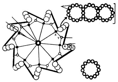
Cart-wheel structure
central rod (HUB)
9-
Spokes
13
Globular subunits
250Å
3
Subtubules (Subfibres)
A-C Connective (DM)
3
Subtubules (Subfibres)
Globular
subunits
(45Å)
250
Å
C
A
B
90o to each other. Each centriole is a microtubular structure and is formed of microtubules arranged in 9 + 0 manner (all the 9 microtubules are peripheral in position). Fig : (A) T.S. Centriole (B) Three subtubules (C) A subtubule Each microtubule is a triplet and is formed of three subtubules which are called A, B and C. A subtubule is about 45Å thick and is formed of 13 parallel protofilaments while each of B and C subtubule is formed of 10 parallel protofilaments. Each protofilament is formed of a row of α, β-tubulin dimers. C sub-tubule of each microtubule is linked to A sub-tubule of adjacent microtubule by a dense material (DM) strand called A-C linker, so all the microtubules are tilted at 40o . Each microtubule is about 250Å in diameter.
Inside the microtubules, there is an intra-centriolar or cart-wheel structure which is formed of a central hub (about 25Å in diameter) and 9 radial spokes or radial fibres. Each radial spoke ends into a dense material (DM) thickening, called X-body or foot which is further linked to A-subtubule. Between two adjacent X-bodies there is another DM-thickening, called Y-body, which is linked to X-body on either side and to A-C linker on outer side.
Centriole is rich in tubulin and ATPase. Centriole can replicate but has no DNA. Centrioles replicate in G2 phase of interphase of cell cycle but do not initiate cell division.
- Chemical composition : Centrosome is lipoproteinaceous structure. The microtubules of centriole are composed of protein tubulin and some lipids. They are rich in ATPase enzyme.
- Origin : The daughter centriole is formed from the pre-existing centriole in G2 of interphase so called selfreplicating organelle.
(6) Functions
- The centrioles help organising the spindle fibres and astral rays during cell division. Therefore, they are called microtubules organising centres. The cells of higher plants lack centrioles and still form a spindle.
- They provide basal bodies which give rise to cilia and flagella.
- The distal centriole of a spermatozoan give rise to the axial filament of the tail.
Important Tips
- Centriole is also called microcentrum or cell centre.
- Each centriole is formed of 9 × 3 = 27 subtubules or subfibres.
| Cilia and flagella. |
- Discovery : Flagellum presence was first reported by Englemann (1868). Jansen (1887) was first scientist to report the structure of sperm flagellum.
- Definition : Cilia and flagella are microscopic, hair or thread-like motile structures present extra-cellularly but originate intra-cellularly from the basal body and help in movements, locomotion, feeding, circulation etc.
- Occurrence : Cilia are found in all the ciliate protozoans e.g., Paramecium, Vorticella etc. flame cells of flat worms; in some larval forms e.g., Trochophore larva of Nereis, Bipinnaria larva of starfish etc.; in some body structures e.g. wind-pipe, fallopian tubes, kidney-nephrons etc.
Flagella are found in all the flagellate protozoans e.g., Euglena, Trichonympha etc., collar cells of sponges; gastrodermal cells of coelenterates; spermatozoa of animals and lower plants; zoospores of algae etc. These are absent in red algae, blue-green algae, angiosperms, nematodes, arthropodes etc.
- Flagella are 1 – 4 per cell where as cilia are infinity in number.
- Cilia are smaller and flagella are longer in size, 5 – 10µmand 150µmrespectively.
- Structure : Both cilia flagella are structurally similar and possess similar parts-basal body, rootlets, basal plate and shaft
(i) Basal body : These are also termed as blepharoplast Membrane
| Radial spoke | |
| (iii) Basal plate : Central fibril develop in this area. It is highly dense and lie above plasma-membrane. | Fig : A diagram of T.S. Cilium or flagellum |
(kinetosome) or basal granule. It is present below the plasma
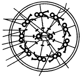
Link
Central
sheath
Bridge
Central
microtubule
membrane in cytoplasm. The structure is similar to centriole made of Outer
9 triplets of microtubules. Out of the 3 fibrils of a triplet first is A microtubules
Inner arm which is round and other two B and C are semi-circular. 9 triplets are Outer arm connected to the centre by spokes. ‘C’ fibrils disappears as it enters Spoke Head into shaft. Interdoublet
link
Subtubule A
(ii) Rootlets : Made of microfilament and providing support to
Subtubule B the basal body. These are striated fibrillar outgrowths.
(iv) Shaft : It is the hair like projecting part of cilia and flagella which remains outside the cytoplasm. It has 9 duplets of microtubules in radial symmetry. These are called axonema. Each axonema has 11 fibrils, 9 in the periphery and 2 in the centre. The arrangement is called 9 + 2 pattern. Central fibrils are singlet fibrils and covered by a central sheath. 9 pheripheral fibrils are duplet and are present at 10o difference from each other. Inner fibril of duplet is known as subfibre A with two bent arms and the outer one is subfibre-B. Peripheral fibrils are linked with each other by peripheral linkage and with the central fibril by radial linkage.
- Chemical composition : Chemically, the central tubules are formed of dynein protein while the peripheral microtubules are formed of tubulin protein. Dynein is the ATPase enzyme which hydrolyses the ATP to provide free energy for ciliary /flagellar beating. The interdoublet linkers are formed of nexin protein. Quantitatively, it is formed of
Proteins = 74 – 84% Lipids = 13 – 23%
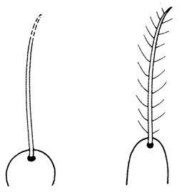 Carbohydrates = 1 – 6% Nucleotides = 0.2 – 0.4%
- Type of flagella : There are two types of flagella.
- Tinsel – type : In this, flagellum has lateral hair-like processes, called flimmers or mastigonemes.
- Whiplash – type : In this, flagellum has no flimmers.
- Motion : Cilia beat in coordinated rhythm either simultaneously (synchronus) or one after the other (metachronic rhythm). The cilia produce a sweeping or pendular stroke. The flagella beat independently, hence produce Whiplash Tinsel
undulatory motion. Fig : Types of flagella
(10) Function
- They help in locomotion, respiration, cleaning, circulation, feeding, etc.
- Being protoplasmic structure they can function as sensory organs.
- They show sensitivity to changes in light, temperature and contact.
- Ciliated larvae take part in dispersal of the species.
- The cilia of respiratory tract remove solid particles from it. Long term smoking damages the ciliated epithelium, allowing dust and smoke particles to enter the long alveoli.
- The cilia of urinary and genital tracts drive out urine and gametes.
Difference between cilia and flagella
| Characters | Cilia | Flagella | ||||
| Number | More in number (may be upto 14,000 per cell). | Less in number (1-8). | ||||
| Size | ||||||
| Small sized (5-10 | µm | ). | Large sized (upto 100-200 | µm | ). | |
| Distribution | Generally distributed on whole body. | Generally located at anterior end of body. | ||||
| Beating | Beat in either metachronous or synchronous coordination. | Beat independently. | ||||
| Type of motion | Sweeping or rowing motion. | Undulatory motion. | ||||
| Function | Locomotion, feeding, circulation, etc. | Only locomotion. | ||||
Important Tips
- Kinocilia : True or motile cilia e.g. of epithelial cells of respiratory tracct.
- Stereo cilia : Immobile cilia e.g. of epididymis.
- Bacterial flagellum consists of a single fibril composed of flagellin protein.
| Cytoskeleton. |
In eukaryotic cell, a framework of fibrous protein elements became necessary to support the extensive system of membranes. These elements collectively form cytoskeleton of the cell. There are of three types.
(1) Microtubules :
-
- Discovery : These were first discovered by De Robertis and Franchi (1953) in the axons of medullated nerve fibres and were named neurotubules.
- Position : The microtubules are electron-microscopic structures found only in the eukaryotic cellular structures like cilia, flagella, centriole, basal-body, astral fibres, spindle fibres, sperms tail, neuraxis of nerve fibres etc. These are absent from amoebae, slime-moulds and prokaryotes.
- Structure : A microtubule is a hollow cylindrical structure of about 250 Å in diameter with about 150 Å luman. Its wall is about 50Å thick. Its walls is formed of 13 parallel, proto-tubules, each being formed of a liner series of globular dimeric protein molecules.
- Chemical composition : These are mainly formed of tubulin protein. A tubulin protein is formed of 2 sub-units : α−tubulin molecule and β−tubulin molecule which are alternatively in a helical manner.
(v) Function
- These form a part of cytoskeleton and help in cell-shape and mechanical support.
- The microtubules of cilia and flagella help in locomotion and feeding.
- The microtubules of asters and spindle fibres of the mitotic apparatus help in the movement of chromosomes towards the opposite poles in cell-division.
- These help in distribution of pigment in the chromatophores, so help in skin colouration.
- These also form micro-circulatory system of the cell which helps in intracellular transport.
- These control the orientation of cellulose microfibrils of the cell wall of plants.
(2) Microfilament
- Position : These are electron-microscopic, long, narrow, cylindrical, non-contractile and proteins structures found only in the eukaryotic cytoplasm. These are present in the microvilli, muscle fibres (called myofilaments) etc. But these are absent from the prokaryotes. These are also associated with the pseudopodia, plasma membrane of fibroblats, etc. These are either scattered or organized into network or parallel arrays in the cytoplasmic matrix.
- Discovery : These were discovered by Paleviz et. al. (1974).
- Structure : Each microfilament is a solid filament of 50-60 Å diameter and is formed of a helical series of globular protein molecules. These are generally grouped to form bundles.
- Chemical composition : These are mainly formed of actin-protein. (v) Functions
- The microfilaments forms a part of cytoskeleton to support the relatively fluid matrix.
- The microfilaments bring about directed movements of particles and organelles along them in the cell.
- The microfilaments also produce streaming movements of cytoplasm.
- The microfilaments also cause cleavage of animal cells which is brought about by contraction of a ring of microfilaments.
- The microfilaments also participate in gliding amoeboid motion shown by amoebae, leucocytes and macrophages.
- The microfilaments are also resoponsible for the change in cell shape curing development, motility and division.
- Myofilaments bring about muscle contraction.
- The microfilaments cause movements of villi to quicken absorption of food.
- The microfilaments are responsible for the movement of cell membrane during endocytosis and exocytosis.
- The microfilaments cause plasma membrane undulations that enable the firoblasts to move.
(3) Intermediate filaments
- Location : They are supportive elements in the cytoplasm of the eukaryotic cells, except the plant cells. They are missing in mammalian RBCes and in the prokaryotes.
- Structure : The IFs are somewhat larger than the microfilaments and are about 10 nm thick. They are solid, unbranched and composed of nonmotile structural proteins, such as keratin, desmine, vimentin.
(iii) Functions
- They form a part of cytoskeleton that supports the fuild cytosol and maintains the shape of the cell.
- They stabilize the epithelia by binding to the spot desmosomes.
- They form major structural proteins of skin and hair.
- They integrate the muscle cell components into a functional unit.
- They provided strength to the axons.
- They keep nucleus and other organelles in place.
Differences between microtubules and microfilaments
| Microtubules | Microfilaments |
| Are hollow cylinders. | Are solid rods. |
| About 200 to 270 Å thick. | About 50 to 60 Å thick. |
| Composed of 13 longitudinal protofilaments each. | Not composed of protofilaments. |
| Formed of protein tubulin. | Formed of proteins actin and myosin. |
| Subunits are dimers that have bound GTP and GDP. | Subunits are monomers that have bound ATP and ADP. |
| Are noncontractile. | Are contractile. |
| Have no role in cytoplasmic streaming, endocytosis and exocytosis. | Play a role in cytoplasmic streaming, endocytosis and exocytosis. |
Important Tips
- Microtubule term was given by Slautterback.
- Tubulin proteins is dimeric protein formed of two globular polypeptides called α− tubulin and β− tubulin.
- Microtubules associated proteins like Tau- protein and kinase control polymerization of tubulin dimer’s.
- Hyman (1917) proposed sol-gel-theory for amoeboid locomotion and was supported by Mast.
| Metabolically inactive cell inclusions/Deutoplasmic substances/Ergastic material . |
Within the cytoplasm of a cell there occur many different kinds of non-living structures which are called inclusions or ergastic substances. They are formed as a result of metabolic activities. They are of following types:
(1) Vacuoles : It is a non-living reservoir, bounded by a differentially or selectively permeable membrane, the tonoplast. The structure of tonoplast is similar to that of single unit membrane i.e. tripartite structure. The vacuole is filled with cell sap or tonoplasm. The thin layer of protoplasm, pushed towards the wall of the cell is called as primordial utricle. They contain water and minerals.
The vacuole in plants was discovered by Spallanzani. The vacuole is not air filled cavity, rather it is filled with a highly concentrated solution the vacuolar sap. It is generally neutral, but at maturity it becomes acidic. The cell sap contains following.
- Gases : CO2, O2 and N2.
- Inorganic salts : Nitrates, chlorides, sulphates, phosphates of K, Na Ca, and Mg.
- Organic acids : Malic acid, formic acid, acetic acid, oxalic acid or their salts.
- Sugars : Cane sugar, glucose and maltose.
- Soluble proteins : Enzymes.
- Glycosides : Like anthocyanins (water soluble pigment)
Some protozoans have contractile vacuoles which enlarge by accumulation of fluid or collapse by expelling them from the cell. The vacuoles may be sap vacuoles, contractile vacuoles or gas vacuoles (pseudo vacuoles).
- Function of vacuoles : Vacuole maintains osmotic relation of cell which is helpful in absorption of water. They also act as reservoir of cells. Turgidity and flaccid stages of a cell are due to the concentrations of sap in the vacuole. In animal cell, it is phagocytic, food vacuole, autophagic or contractile in nature.
(2) Reserve food material
The reserve food material may be classified as follows :–
Food
Nitrogenous Non-nitrogenous
Proteins
Amides
Fats and oils
Carbohydrates
Starch Cellulose Sugar Glycogen Inulin
(i) Carbohydrates : Non-nitrogenous, soluble or non- soluble important reserve food material. Starch cellulose and glycogen are all insoluble.
- Starch : Found in plants in the form of minute solid grains. Starch grains are of two types:
Assimilation starch : It is formed as a result of photosynthesis of chloroplasts. Diastase enzyme converts it into soluble sugar at night time. The conversion of sugar into reserve or storage starch is brought about by leucoplast as amyloplast.
Reserve starch : Thick layers are deposited around an organic centre called hilum. When hilum is situated just at the centre of starch grain, it is said to be concentric e.g. pea, bean, wheat etc. While it is situated not at the centre, but nearer the margin it is said to eccentric e.g. potato.
- Glycogen : Glycogen or animal starch occurs only in colourless plants like fungi. It occurs in the cytoplasm as an amorphous body.
- Inulin : It is a complex type of polysaccharide, soluble found dissolved in cell sap of roots of Dahlia, Jaruslem, Artichoke, Dandelion and members of compositae. When these roots are preserved in alcohol it precipitates in the form of “ Sphaerites” or fan shaped crystals.
- Sugars : A number of sugars are found in solution of cell sap. These include glucose, fructose, sucrose, etc. Glucose and fructose are monosaccharides while can sugar is disaccharide and occurs in beet root and sugar- cane.
- Cellulose : Chemical formula is (C H O6 10 5)n. The cell wall is made up of cellulose. It is insoluble in water.
- Fats and Oils : These are important reserve food material. These are always decomposed into glycerol and fatty acids by enzymatic action. Fat is usually abundant in cotyledons than in the endosperm. e.g. flax seed produce linseed oil, castor produce castor oil, cotton seeds produce cottonseed oil, etc.
- Proteins and Amides (Aleurone grains) : Storage organ usually contain protein in the form of crystalline bodies known as crystalloids (potato). Proteins may be in the form of aleurone grains as in pea, maize, castor, wheat, etc. Each aleurone grain consists of a large crystalline grain of protein known as crystalloid associated with it there is a smaller body globoid. It is not a protein but double phosphate of calcium and magnesium.
(3) Excretory Products : The organic waste products of plants are by-product of metabolism. They are stored as inclusions. Depending upon chemical composition they are classified as:
- Resins : They are believed to be aromatic compounds consisting of carbon, hydrogen and oxygen and are acidic in nature. Sometimes they are found in combination with gums and are called gum resin. e.g. Asafoetida (heeng). These are used in making varnishes and gums.
- Tannins : They are complex nitrogenous compounds of acid nature having an astringent taste. They are used in conversion of hide into leather. With ferric salt they are largely used manufacture of ink. Presence of tannin in plants makes its wood hard durable and germ proof.
- Alkaloids : These are organic, basic, nitrogenous substance. They occur in combination with organic acids and most of them are poisonous. From plants, cocaine, hyoscine, morphine, nicotine, quinine, atropine, strychnine and daturine etc. are extracted.
- Glucosides : Some glucosides or glycosides function as storage substance e.g. amygdaline of the bitter almond. Erythrocyanins and Anthocyanins are responsible red and blue colour and flavines for cream colour. Carotene is an unsaturated fatty acid and not a glycoside, gives red and orange colour to roots.
- Etherial and Essential oils : These consist mixture of various hydrocarbons known as tarpenes and their oxygen derivatives. They are responsible for flavor of many fruits and scent of many flowers etc. They are volatile and are soluble in water, ether, petroleum etc. e.g. lavender, mint, clove oil, eucalyptus oil, theme oil etc.
- Mineral matter : Many minerals are waste products in plants.
- Calcium oxalate : It occurs in the form of crystals of various shapes.
Raphides : Needle shaped crystals are known as raphides. They are found single or in bundles. e.g. in plants like jamikand, Colocasia, water hyacinth (Jal kumbhi), amorphophallus and aroids.
Rosette or Sphaeraphides : Star shaped crystals. They occur in special mucilaginous parenchyma cells of the petiole of arum, water hyacinth, etc. Crystals in the form of cubes are found in tunic of onion bulb. In the leaf of belladona, these crystals are in the form of sand and also called as sand crystals.
Calcium oxalate crystals : In members of family solanaceae. They are found as cubics, rods and prisms.
- Calcium carbonate : It is deposited in the form of crystalline masses hanging from a cellulose stalk in enlarged epidermal cells of leaves of Ficus elastica (Indian rubber plant) and is called as cystolith.
- Latex : It is an emulsion in water having many substances either in suspension or in true solution. It may contain sugars, alkaloids and oils. It is watery in banana, milky white in Euphorbia, yellow or orange red in opium (poppy) is dried latex.
- Organic acids : Tartaric acid in tamarind, and grapes, citric acid in lemon, orange etc. malic acid in apple and Bryophyllum. Oxalic acid in the form of crystals.
- Gums : It is formed by decomposition of cellulose cell wall. Gum arabic of commerce is obtained from Acacia senegal.
(4) Secretory products : The chief secretion of plants are enzymes nectar, colouring matter, water etc. These secretion are helpful to plants.
| Nucleus . |
- Definition : (Karyon = Nucleus) The nucleus also called director of the cell. It is the most important part of the cell which directs and controls all the cellular function.
- Discovery : The nucleus was first observed by Robert Brown (1831). Nucleus plays determinative (in heredity) role in cell and organism, that was experimentally demonstrated by Hammerling (1934) by conducting surgical experiments with green marine unicelled algae Acetabularia.
- Occurence : A true nucleus with definite nuclear membrane and linear chromosome, is present in all the eukaryotes except mature mammalian RBCs, sieve tube cell of phloem, tracheids and vessels of xylem. The prokaryotes have an incipient nucleus, called nucleoid or prokaryon or genophore or false nucleus or bacterial chromosome.
- Number : Usually there is a single nucleus per cell i.e. mononucleate condition, e.g. Acetabularia.
- Anucleate (without nucleus) : RBCs of mammals, phloem sieve tube, trachids and vessels of xylam.
- Binucleate : e.g. Ciliate, Protozoans like Paramoecium.
- Polynucleate : e.g. fungal hyphae of Rhizopus, Vaucheria. Polynucleate condition may be because of fusion of a number of cells. i.e. syncytium, coconut endosperm or by free nuclear divisions without cytokinesis i.e. coenocyte.
- Shape : It varies widely, generally spherical e.g. cuboidal germ cells, oval e.g. columnar cells of intestine, bean shaped in paramoecium, horse-shoe shaped in vorticella, bilobed, e.g. WBCs (acidophils), 3 lobed e.g. basophil, multilobed e.g. neutrophils, long and beaded form (moniliform) e.g. stentor and branched in silk spinning cells of platy phalyx insect larva.
- Size : The size of nucleus is variable i.e. 5 – 30µ. In metabolically active cells size of the nucleus is larger than metabolically inactive cells. The size depends upon metabolic activity of the cells. It is directly proportional to number of chromosomes.
(7) Chemical composition of nucleus
Proteins = 80% (65% acidic, neutral and enzymatic proteins; 15% basic proteins-histones)
DNA = 12%
RNA = 5%
Lipids = 3%
Enzymes like polymerases are abundantly present and help in synthesis of DNA and RNA. Minerals like Ca2+,Mg2+,Na+, and K+ are present in traces.
(8) Ultrastructure : The nucleus is composed of following structure
(i) The nuclear membrane (ii) The nucleous.
- The nuclear sap or nucleoplasm.
- The chromatin fibres.
The nuclear membrane or karyotheca
- Definition : It is defined as a regulatory envelope which controls the nucleo-cytoplasmic interacitons and
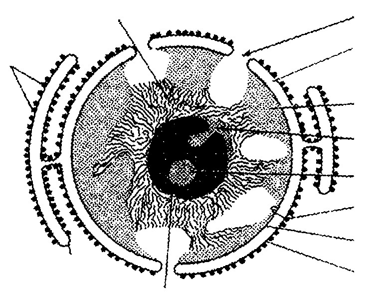
Endoplasmic
reticulum
Nucleolus
Euchromatin
exchange of materials.
Nuclear pore
- Discovery : Nuclear membrane, also called Parinuclear space
Ribosomes nuclear envelope or nucleolemma or karyotheca, was
first discovered by Erclab (1845). Heterochromatin
Perinucleolar
- Structure : It is a bilayered envelope. Each chromatin membrane is about 90 Å thick lipoproteinous and Intranucleolar chromatin trilaminar. Outer membrane, called ectokaryotheca, is Nuclear envelope studded with ribosomes on its cytoplasmic surface and Inner membrane is continuos with RER at some points. Inner membrane, Outer membrane called endokaryotheca, is without ribosomes and is
Fig : Electron microscopic structure of nucleus internally lined by electron-dense material of protein fibres called fibrous or nuclear lamina nuclear cortex or hoeny comb layer (about 300 Å thick). Two membranes are separated by a fluid-filled intermembranous perinuclear space (about 100-300Å). Nuclear membrane contains following structure.
- Nuclear pore : Nuclear membrane is porous and has 1,000-10,000 octagonal nuclear pores. Each nuclear pore is about 400-1,000 Å in diameter (average size is 800 Å). The number and size of the nuclear pores depend upon the needs of the cell.
Nuclear pores are interspaced at about Outer nuclear membrane Pore complex Ribosomes Perinuclear space
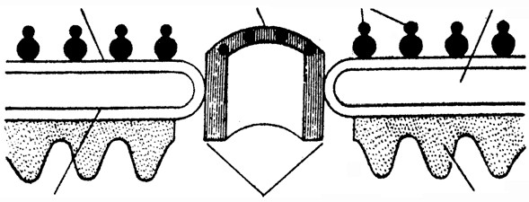
Fibrous lamina
1000-1500 Å. Each nuclear pore is fitted with a cylindrical structure, called annulus (with a lumen of 500 Å) and both collectively form the pore complex or pore basket. Annulus has 8 microcylinders (each about 200 Å in diameter and with a lumen of 50 Å) in its wall. It Inner nuclear membrane Annulus
also encloses a channel having Fig : V.S. of nuclear envelope showing nuclear pore, Ribosomes and fibrous lamina nucleoplasmin for the movement of substances. Annulus acts as a diphragm and regulates the size of the nuclear pore.
- Nuclear blebbing : The nuclear envelope shows evagination. As a result, blebs are formed which are pinched off. This phenomenon is called blebbing. The nuclear vesicles so formed are thought to give rise to mitochondria, plastids, etc. Blebbing may also occur from the outer unit membrane only. A row of these blebs move towards the periphery. As a result of deposition of matrix material in between these blebs, and annulate lamella is formed. The annulate lamellae is thought to give rise to ER cisternae.
(iv) Origin : It is formed by the fusion of ER elements during the telophase of cell division. (v) Functions
- It regulates the nucleo-cytoplasmic interactions.
- It allows the passage of inorganic ions and small organic molecules.
- It helps in pinocytosis and phagocytosis of large sized molecules .
- It allows passage of ribosomal subunits, RNAs and proteins through nuclear pores.
- It maintains the shape of the nucleus.
- Fibrous lamina strengthens the nuclear envelope. It also helps in dissolution and reformation of nuclear membrane during cell division.
The nucleolus (Little nucleus)
- Discovery : Nucleolus was first observed by Fontana (1781) in the skin cells of an eel. Term ‘nucleous’ was coined by Bowman (1840). Its light microscopic structure was given by Wagner (1840).
- Position : It is generally associated with nucleolar organizer region (NOR) of the nucleolar chromosomes. It is absent in muscle fibres, RBC, yeast, sperm and prokaryotes.
- Number : Generally, a diploid cell is with two nucleoli but there are five nucleoli in somatic cell of man and about 1000 nucleoli in the oocytes of Xenopus.
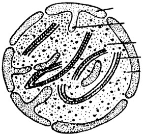 Structure : (De Robertis et. al 1971). A nucleolus is Perinucleolar distinguishable into following regions :- chromatin
Intranucleolar
- Chromatin : The nucleolus is surrounded by perinucleolar chromatin
Matrix chromatin. Heterochromatic intrunsions are also seen in the nucleolus Fibrils
which constitutes the intranucleolar chromatin. Granules
- Pars fibrosa : Fibrils of 80 – 100 Å size form a part of the nucleolus.
- Pars granulosa : Granules of 150 – 200 Å diameter constitute the granular part of the nucleolus. They appear like vesicle with a light central core. The granules may be joined by filament forming a beaded Fig : Ultrastructure of a Nucleolus primary nucleolonema. The fibrils may also be associated to it. The primary nucleolonema may further coil to form the secondary nucleolonema.
- Pars amorpha : The granules and the fibrils lie dispered in an amorphous proteinaceous matrix. Nucleolus contains large amount of proteins mainly phosphoproteins. There are no histones proteins. RNA methylase, an enzyme that transfers methyl groups to the RNA bases has been localized in nucleolus. Nucleolus is stained by “pyronine”. It is not bounded by any limiting membrane. Fibrillar region of nucleolus is called secondary constriction or nucleolar organising region (NOR) and this region directs the synthesis of rRNA. Ribosomes are assembled here as such it is also called ribosome producing machine or factory. Ribosomal units so formed are joined together by thin filament (rRNA) forming a structure like string of beads and it is called “nucleonema”.
- Chemical composition : Nucleolus is mainly formed of RNA and non histone acidic proteins. It is a storehouse of RNA.
- Origin : A nucleolus is formed at specific sites, called the nucleolar organizers, present on certain chromosomes region (NOR).
(vii) Functions
- It is seat of biogenesis of rRNA and also stores rRNA.
- It plays important role in spindle formation during cell division.
- It receives the ribosomal proteins from the cytoplasm, combines the rRNAs and ribosomal proteins to form ribosomal subunits.
Nucleoplasm : It is also called karyolymph. It is transparent, homogenous, semifluid, colloidal, ground substance present inside the nuclear membrane in which nuclear chromatin and nucleoli are embedded. Chemically it contains. Nucleoplasm is also known as protoplasm of nucleus.
- Nucleic acid : Monomer nucleotides of DNA and RNA
- Proteins : Basic proteins (nuclear protamines and nucleohistones and acidic proteins (non-histone)
- Enzymes : DNA polymerase, RNA polymerase, NAD synthetase, nucleoside triphosphatase, and pyruvic acid kinase, etc.
- Minerals : Phosphorus, potassium, sodium, calcium, magnesium, etc.
- Ribonucleoproteins : Contain perichromatin granules and interchromatin granules. Histone proteins are basic because they contain arginine in much amount e.g. H1,H A H B H2 , 2 , 3 and H4 .
The nucleoplasm helps in maintaining the shape of nucleus formation of spindle protein of NAD, ATP, DNA, RNAs and ribosomal subunits. Plasmosome and karyosome combindly called “amphinucleoli”.
Chromatin fibres /Nuclear chromatin : The nucleoplasm contains many thread like, coiled and much elongated structures which take readily the basic stains such as “basic fuschin”. These thread like structures are known as chromatin fibre. They are uniformly distributed in the nucleoplasm. They are observed only in the “interphase stage”. Chromatin fibres are made of chromosomes. In resting nondividing eukaryotic cells the genome is nucleoprotein complex and it is called chromatin.
| Chromosome . |
- Definition : During interphase, chromatin threads are present in the form of a network called chromatin reticulum. At the time of cell division, these thread like structures of chromatin become visible as independent structures, called chromosomes.
- Structure of chromosome : Each chromosome consists of two coiled filaments throughout its length called chromonemata by Vejdovsky. These have bead like structures called chromomeres which bear genes. Chromatid is a half chromosome or daughter chromosome. The two chromatids are connected at the centromere or primary constriction. Primary constriction (centromere) and secondary constriction gives rise to satellite. The secondary constriction consists of genes which code for ribosomal RNA and nucleolus hence it is called as “nucleolar organizer region”. Chromosomes having satellite are called SAT chromosomes.
The ends of chromosomes are called “telomeres” (which do not unite with any other structure). A tertiary constriction is also present in chromosomes, which perhaps helps in recognition of chromosomes.
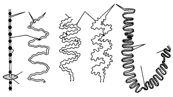
Chromonemata
Telameresa
Chromonema
Telomeres
Chromonemata
Satellite
Centromere
Nucleolus
Nucleolar
organizers
Chromomeres
Centromers
A
B
C
D
E
In 1928 Emile Heitz developed a technique for stainning of chromosomes. Chromosomes can be stainned with acetocarmine or fuelgen (basic fuschin) there are two types of regions are seen :–
(i) Heterochromatin : It is formed of thick regions which are more darkly stained than others
Fig : Chromosomes A. Diagrammatic B, C, D, E-Different
areas. It is with condensed DNA which is parts of chromosome transcriptionally inactive and late replicating. It generally lies near the nuclear lamina. Heterochromatin are of two types : –
- Facultative heterochromatin : Temporarily inactivated chromatin and forms 2.5% of the genome.
- Constitutive hetrochromatin : Permanently inactivated chromatin and generally ground near centromeres.
(ii) Euchromatin : It is true chromatin and is formed of thin, less darkly stained areas. It is with loose DNA which is transcriptionally active and early replicating.
(3) Chemical chomposition : DNA – 40%. Histone – 50%. Other (acid) Proteins – 8.5%. RNA – 1.5%. Traces of lipids, Ca, Mg and Fe. Histone are low molecular weight basic proteins which occur alongwith DNA in 1:1ratio. Nonhistone chromosomal or NHC proteins are of three types– structural, enzymatic and regulatory. Structural NHC proteins form the core or axis of the chromosome. They are also called scaffold proteins. Enzymatic proteins form enzymes for chemical transformation, e.g., phosphates, RNA polymerase, DNA polymerase. Regulatory proteins control gene expression. HMG (high mobility group) proteins get linked to histones for releasing DNA to express itself.
(4) Ultrastructure and Models of chromosomes : (See in genetics). Important Tips
- Syncytium is multinucleate condition formed by the fusion of cells e.g. in plasmodium of slime moulds.
- Coenocytic is multinucleate condition by repeated Karyokinesis but not followed by cytokinesis e.g. in vaucheria, rhizopus.
- Callan and Tamlin (1950) first to observe nuclear pore in nuclear membrane.
- Staining property of chromosomes is called as heteropycnosis.
- Satellite is also called trabant.
- Centromere or kinetochore is responsible for chromosomal movement during cell division.
- Idiogram : Karyotype of a species is represented with the help of a diagram called idiogram.
- Genome : It is defined as the haploid set of chromosomes.
- Plasmon : Genes present in cytoplasm are called “plasmons”
- Non histone proteins (acidic proteins) are rich in nucleus and less chromosome.
| Micromolecules . |
- Definition : These are molecules of low molecular weight and have higher solubility. These include minerals, water, amino acid, sugars and nucleotides. All molecules or chemicals functional in life activity are called biomolecules.
- Elements : They are naturally occuring and they are classified on the basis of their property into metals and non-metals. Again on the basis of presence and requirement in plants and animals, they are grouped into major and minor bioelements. Which are required in large amount are major bioelements e.g. Ca, P, Na, Mg, S, K, N, etc., while those are required in small amount are called minor bioelements e.g. Fe, Cu, Co, Mn, Mo, Zn, I, etc.
On the basis of function, they may be of following types :– (i) Framework elements : Carbon, oxygen and hydrogen.
- Protoplasmic elements : Protein, nucleic acid, lipids, chlorophyll, enzymes, etc.
- Balancing elements : Ca, Mg and K. counteract the toxic effect of other minerals by ion-balancing.
There are 17 essential elements in plants and 24 in animals. 14 elements are non-essential :– (iv) Proportion of elements in a cell
| Oxygen – O – 62% | Chlorine- Cl | 0.16% |
| Carbon – C– 20% major elements (95%) | Sulphur – S | 0.14% |
| Hydrogen- H– 10% | Potassium- K | 0.11% |
| Trace elements- 0.75% minor elements (4.25%) | Sodium – Na | 0.10% |
| Calcium – Ca- 2.5% | Magnesium – Mg | 0.07% |
| Phosphorous- P- 1.14% | Iodine- I | 0.14% |
| Iron – Fe | 0.10% |
(3) Biological compounds : These involve two kinds of compounds.
- Inorganic compounds : Characterised by absence of carbon, simple structure with low molecular weights
e.g. water, minerals, ions and gases etc. Water 80% and inorganic salts 1-3%.
- Organic compounds : Characterised by presence of carbon bonded to form a straight chain or ring structure.
| Carbohydrates | Lipids | Proteins | Nucleotides | Other compounds |
| 1.0% | 3.5% | 12.0% | 2.0% | 0.5% |
- Cellular pool : Aggregated and interlinked various kinds of biomolecules in a living system. So cell is called cellular pool. It includes over 5000 chemicals. Inorganic chemicals are present mostly in aqueous phase while organic in both. The aqueous phase may be moleculer solution in which dissolved particles are smaller than 0.000001 mm and colloidal phase in which particle size varies between 0.0001 – 0.000001 mm. Cellular pool comprises of both crystelloid and colloidal particles. Hence called as crystal colloids the non-aqueous phase comprises of organic molecules present in cell compartments like plasma membrane, mitochondria, chloroplast, etc.
- Water : Liquid of life, major constituent of cell (about 60-90%) and exists in intracellular, intercellular and in vacuoles. In cells it occurs in free state or bound state (KOH, CaOH etc.).
(i) Properties of water : It is colourless, transparent, tastless and odourless, neutral (pH-7) liquid. It is universal solvent, as it can dissolve both polar and non-polar solutes. High boiling point due to hydrogen bonding. Shows high degree of cohesion and adhesion. It can undergo three states of matter i.e. solid liquid gas. It is dense and heaviest at 4C and solid below it.
(6) Carbohydrates : e.g. sugars, glycogen (animal starch), plant starch and cellulose.
- Source of carbohydrate : Mainly photosynthesis. It exists only in 1% but constitutes 80% of the dry weight of plants.
- Composition : It consists of carbon, hydrogen and oxygen in the ratio C Hn 2n nO . It is also called saccharide and sugars are their basic components. Classification of carbohydrates can be summarised as :–
Carbohydrates
Monosaccharides
e.g.
Monosaccharides and
their derivatives
Oligosaccharides (number of
monosaccharides from 2 to 10, di,
tri, tetrasaccharides etc.)
Polysaccharides (number of
monosaccharides over 10)
Derivatives of
monosaccharides
Heteropolysaccharides
(
glycoproteins, starch,
Homopolysaccharides
starch, amylopectin,
(
glycogen, cellulose etc.)
Triose – 3C, Tetrose – 4
C
Pentose – 5C, Hexose – 6
C
Heptose – 7C, Octoses – 8
C
Nanoses-9C, Decoses – 10
C
proteins)
| Uric acid e.g.
glycouronic acid, galactouronic acid |
Aroic acids e.g. glucaroic acid, galactaroic acid | Aminosaccharides e.g.
glucosamine, galactosamine |
Phosphosaccharides e.g. Glycosides e.g.
glucose-6- phosphate, nucleotides, nucleosides, fructose 1,6 biphosphate etc. Coenzymes etc. |
Monosaccharides : These are single sugar units which can not be hydrolysed furthur into smaller carbohydrates. General formula is C Hn 2n nO , e.g. Triose-3C, glyceraldehyde, dihydroxyacetone, etc., tetrose, pentose, hexose, etc. About 70 monosaccharides are known, out of which only 20 are present in plants and animals.
(i) Important Hexoses
- Glucose : C H O6 12 6. Grape sugar is dextrose. Grape is sour due to presence of tartaric acid. Fructose is called fruit sugar (sweetest among natural sugars) and glucose is called ” sugar of body”. Normal level of blood glucose is 80-120mg/100ml. If it exceeds then condition is called “glucosuria”.
- Fructose : Occurs naturally in fruit juices and honey. Hydrolysis of cane sugar in body also yields fructose.
- Galactose : It is called as brain sugar. It’s an important constituent of glycolipids and glycoproteins.
- Mannose : It is obtained on hydrolysis of plant mannans and gums. It is constituent of albumins, globulins
and mucoproteins.
(ii) Structure of monosaccharides (iii) Properties of monosaccharide
- Monosaccharides are colourless, sweet tasting, solids.
- Due to asymmetric carbon, they exist in different isomeric forms. They can rotate polarized light hence they are dextrorotatory and leavorotatory.
- D-glucose after reduction gives rise to a mixture of polyhydroxy alcohol, sorbitol or mannitol.
- The sugars with a free aldehyde or ketone group reduce Cu++ to Cu+ (cupric to cuprous)
- Sugars show oxidation, esterification and fermentation.
- The aldehyde or ketone group of a simple sugar can join an alcoholic group of another organic compound bond C-O-C the process involves loss of water and is called condensation (H-O-H) or H+OH→ H O2 .
(iv) Functions of monosaccharides
H
H O
|
H – C – H
C
1| 1|
H – C = OH
H – C – OH
|
|
HO – C – H
HO – C – H
|
|
H – C – OH
H – C – OH
|
|
H – C – OH
H – C – OH
|
|
H – C – OH
H – C – OH
6|
6|
H
H
OH
CH
2
|
C
|
1
H
H
|
C
|
HO
H
|
C
|
OH
OH
|
C
|
H
O
H
|
C
|
HO
2
3
4
5
6
6
OH
CH
2
OH
CH
2
1
|
C
|
HO
H
|
C
|
OH
OH
|
C
|
H
O
H
|
C
|
3
4
5
2
Glucose
Pyranose Form)
(
Fructose
(Pyranose Form)
Fig : Open chain and ring forms of three hexoses
- Glucose is the ultimate source of ATP in the cell respiration.
- It is used in formation of vitamin C.
- The intermediate compounds for the formation of glucose in photosynthesis are triose, tetrose, pentose and heptose, etc.
- Galactose is a constituent of agar-agar.
- Glucose is a blood sugar and xylose is a non nutritive sweetner.
- Polymerisation of these molecules forms macromolecules.
- Ribose and deoxyribose are constituent of nucleic acids and nucleotides (h) Glyceraldehyde and dihydroxyacetone are trioses.
- Sugars have free aldehyde or ketone group which can reduce Cu++ to Cu+ and are called reducing sugars. Benedicts or fehling’s test are used to confirm the presence of reducing sugars.
Oligosaccharides : Formed due to condensation of 2-10 monosaccharide units, the Oxygen bridge is known as “glycoside linkage” and water molecule is eliminated. The bond may be α and β.
C
H
O
C
H
C
C
C
C
H
H
C
O
C
C
C
C
C
H
H
H
H
OH OH OH OH
α-glycosidic linkage β-glycosidic linkage
(i) Disaccharides : Composed of two molecules of same or different monosaccharide units. Also called “double sugars”. Molecular formula is C12 22 11H O .
- Maltose : Also called “malt sugar” stored in germinating seeds of barley, oat, etc. It is formed by enzymatic (enzyme amylase) action on starch. It is a reducing sugar.
- Sucrose : “Cane sugar” or ” table-sugar”. Obtained from sugarcane and beet root and on hydrolysis splits into glucose and fructose.
- Lactose : Milk sugar or 5% in mammalian milk. On hydrolysis yields glucose and galactose. Streptococus lacti converts lactose in to lactic acid and causes souring of milk.
- Trisaccharides : Composed of three molecules of sugars. Molecular formula is C H18 32 16O .
- Raffinose : Found in sugar beet, cotton and in some fungi. It is made up of glucose, fructose and galactose.
- Gentianose : Found in rhizomes of gentian species, made up of glucose and fructose.
- Tetrasaccharides : Composed of four molecules of same or different sugars. Stachyose is found in Stachys tubefera. It is made up of two unit of galactose, one unit of glucose and one unit of fructose.
- Polysaccharides : General formula is (C H6 10 5O )nformed by condensation of several molecules (3001000) of monosaccharides, (Described under ” Macromolecules”).
- Reducing and Non-reducing carbohydrates : Those which reduce Tollen’s reagent or fehling solution are called reducing sugars and those do not reduce are called non-reducing sugars. All monosaccharides and disaccharides except sucrose are reducing. While all polysaccharides are non-reducing sugars.
(7) Lipids : Term lipid was coined by Bloor. These are esters of fatty acids and alcohol. They are hydrophobic insoluble in water but soluble in benzene, ether and chloroform. Lipids are classified into three groups:–
(i) Simple lipids : These are the esters of fatty acids and glycerol. Again they are typed as :–
- Fats and Oils : (Natural lipids or true fats). These triglycerides of fatty acid and glycerol. Fats which are liquid at room temperature are called oils. Oils with polyunsaturated fatty acids are called polyunsaturated e.g. sunflower oil, lower blood cholesterol.
- Fatty acids : Obtained by hydrolysis of fats. Formic acid is simplest fatty acid (HCOOH). These are of 2 types :–
Saturated fatty acids : The fatty acids which do not have double bond in between carbon atoms.e.g.
butyric acid, palmitic acid,hexanoic acid, etc. They have high melting points, solid at room temperature and increase blood cholesterol.
Unsaturated fatty acids : The fatty acids which have double bonds in carbon atoms. e.g. 8 hexadecanoic acid, 9 octadecanoic acid etc. They have lower melting points mostly found in plant fats, liquid at room temperature and lower the blood cholesterol.
- Waxes : These are simple lipids composed of one molecule of long chain fatty acid and long chain monohydric alcohol. Waxes have high melting point, insoluble in water, resistant to atmospheric oxidation, chemically inert and not digested by enzymes. They reduce rate of transpiration by making plant tissue water proof and work as excellent lubricant.
Types of waxes
- Plant wax : Forms coating.
- Bee’s wax : It is secretion of abdominal glands of worker honeybee. It consist of palmitic acid and myricyl alcohol.
- Lanolin or Wool fat : It is secreted by cutaneous glands, also obtained from wool of sheeps. It consists of palmitic acid, oleic or stearic acid and cholesterol.
- Sebum : It is secretion of sebaceous gland of skin.
- Paraffin wax : Obtained from petrolium.
(ii) Compound lipids : They contain some additional or element. Group with fatty acid and alcohol on the basis of group they may be of following types:
- Phospholipids : These contain phosphoric acid. It helps in transport, metabolism, blood clotting and permeability of cell membrane. It is a bipolar molecule i.e. phosphate containing end is hydrophilic whereas fatty acid molecules represent hydrophobic (non-polar tail). Phospholipids again comprises.
Lecithin : These are yellowish grey solids, soluble in ether and alcohol but insoluble in acetone. On hydrolysis they yield glycerol, fatty acid, phosphoric acid and choline. Lecithins are broken down by enzyme lecithinase to lysolecithin. The enzyme is found in venom of bee and cobra.
Cephalins : Found in animal tissue and soyabean oil. Cephalin contains choline or serine sometimes and stearic acid, oleic acid, linoelic and arachidonic acid.
- Glycolipids : These contain nitrogen and carbohydrate beside fatty acids. Generally found in white matter of nervous system. e.g. sesocine frenocin.
- Chromolipids : It includes pigmented lipids e.g. carotene.
- Aminolipids : Also known as sulpholipids. It contains sulphur and amino acids with fatty acid and glycerol. Cutin and suberin are also compound lipids resistant to water and also provide mechanical support in plants.
(iii) Derived lipids : These are obtained by hydrolysis of simple and compound lipids. Derived lipids include following components :–
- Sterols : Lipids without straight chains are called sterols. They are composed of fused hydrocarbon rings and a long hydrocarbon side chain. Best known sterol is cholesterol, present in high concentration in nervous tissue and in bile. Cholesterol is also the precursor of hormones like progesterone, testosterone, estradiol and cortisol and vitamin D. Diosgenin is obtained from yam plant (Dioscorea) used in making anti- infertility pills.
- Digitalin : It is prepared from leaves of Foxglove (Digitalis lantana) is a heart stimulant.
- Ergosterol : Present in food, found in ergot and yeast. It is precursor of another form of vitamin D, ergocalciferol (D2).
- Coprosterol : It is found in faeces. It is formed as a result of the reduction by bacteria in intestine from the double bond of cholesterol between C5 and C6.
- Tarpens : It is essential oil and present mostly in oils of camphor, eucalyptus, lemon and mint. Phytol is a terpenoid alcohol present in Vitamin A, K, E and in pigments like chlorophyll carotenoid. Other forms are licopene, gibberellins and natural rubber.
- Prostaglandin : It is hormone like compound derived from arachidonic acid. Mostly present in secretion of seminal vesicles in males and menstrual cycle fluid in females.
- Blubber : A very thick layer of subcutaneous fat in whale. (iv) Functions of lipids
- Oxidation of lipids yields comparatively more energy in the cell than protein and carbohydrates. 1gm of lipids account for 39.1 KJ.
- The oil seeds such as groundnut, mustard, coconut store fats to provide nourishment to embryo during germination.
- They function as structural constituent i.e. all the membrane system of the cell are made up of lipoproteins.
- Amphipathic lipids are emulsifier.
- It works as heat insulator.
- Used in synthesis of hormones.
- Fats provide solubility to vitamins A, D, E, and K.
(8) Amino acids : Amino acids are normal components of cell proteins (called amino acid). They are 20 in number specified in genetic code and universal in viruses, prokaryotes and eukaryotes. Otherwise amino acids may be termed rare amino acids, which take part in protein synthesis e.g. hydroxyproline and non- protein amino acids do not take part in protein synthesis e.g. Ornithin, citrullin, gama-aminobutyric acid (GABA) a neurotransmitter, etc.
(i) Structure and Composition : Amino acids are basic units of protein and made up of C, H, O, N and sometimes S. Amino acids are organic acids with a carboxyl group (–COOH) and one H amino group (−NH2) on the α -carbon atom. Carboxyl group attributes acidic | properties and amino group gives basic ones. In solution, they serve as buffers and help NH2 − −C| COOH to maintain pH. General formula is R − CHNH2.COOH . R
Amino acids are amphoteric or bipolar ions or Zueitter ions. Amino acids link with each other by peptide bond and long chains are called polypeptide chains.
(ii) Classification
Based on R–group of amino acids.
- Simple amino acids : These have no functional group in the side chain. e.g. glycine, alanine , leucine, valine etc.
- Hydroxy amino acids : They have alcohol group in side chain. e.g. threonine, serine, etc.
- Sulphur containing amino acids : They have sulphur atom in side chain. e.g. methionine, cystenine.
- Basic amino acids : They have basic group (−NH2) in side chain. e.g. lysine, arginine.
- Acidic amino acids : They have carboxyl group in side chain. e.g. aspartic acid, glutamic acid.
- Acid amide amino acids : These are the derivatives of acidic amino acids. In this group, one of the carboxyl group has been converted to amide (−CO NH. 2). e.g. asparagine, glutamine.
- Heterocyclic amino acids : These are the amino acids in which the side chain includes a ring involving at least one atom other than carbon. e.g. tryptophan, histidine.
- Aromatic amino acids : They have aromatic group (benzene ring) in the side chain. e.g. phenylalanine, tyrosine, etc.
On the basis of requirements : On the basis of the synthesis amino acids in body and their requirement, they are categorized as :–
- Essential amino acids : These are not synthesized in body hence to be provided in diet e.g. valine, leucine, isoleucine, theronine ,lysine, etc.
- Semi-essential amino acids : Synthesized partially in the body but not at the rate to meet the requirement of individual. e.g., arginine and histidine.
- Non-essential amino acids : These amino acids are derived from carbon skeleton of lipids and carbohydrate metabolism. In humans there are 12 non- essential amino acids e.g. alanine, aspartic acid, cysteine, glutamic acid etc. Proline and hydroxyproline have, NH (imino group) instead of NH2 hence are called imino acids. Tyrosine can be converted into hormone thyroxine and adrenaline and skin pigment melanin. Glycine is necessory for production of heme. Tryptophan is the precursor of vitamin nicotinamide and auxins. If amino group is removed from amino acid it can form glucose and if COOH group is removed, it forms amines e.g. histamine.
(iii) Functions of amino acids
- Amino acids are building blocks of proteins and enzymes.
- By glycogenolysis, they form glucose.
- Hormones like adrenaline and thyroxine are formed with the help of tyrosine.
- Antibiotics often contain non-protein amino acids.
- They are precursour of many substances.
(9) Nucleotides : Structurally a nucleotide can be regarded as a phosphoester of a nucleoside. A combination of nitrogens base and a sugar is called nucleoside and combination of a base, a sugar and phosphate group is known as nucleotide.
| Types of nitrogen base | Nucleoside | Nucleotide |
| Adenine | Adenosine | Adenylic acid |
| Guanine | Guanosine | Guanylic acid |
| Cytosine | Cytidine | Cytidilic acid |
| Thymine | Thymidine | Thymidylic acid |
| Uracil | Uridine | Uridylic acid |
There are two types of pentose sugars, ribose found in RNA and deoxyribose found in DNA. Nucleotides form 2% of the cell component.
N2 base + Pentose sugar→ ‘Nucleoside’
Nucleoside + Phosphoric acid→ ‘Nucleotide’ + H O2 .
There are two types of bases which occur in the nucleic acids.
- Purines : Purines are 9 membered double ringed nitrogenous bases which possess nitrogen at 1′ ,3′ ,7′ and 9′ positions. They are adenine (A) and guanine (G).
- Pyrimidines : They are smaller molecule than purines. These are 6 membered single ringed nitrogenous bases that contain nitrogen at 1′ and 3′ positions like cytosine (C), thymine (T) and uracil (U). In DNA adenine pairs with thymine by two H2 bond and cytosine pairs with guanine by three H bond.
A nucleotide may have one, two or three phosphates, as one in AMP (adenosine monophosphate), two in ADP (adenosine diphosphate). The phosphate bond is called high energy bond and it release about 8 K cal. ATP was discovered by Karl Lohmann (1929). Formation of ATP is endergonic reaciton.
(iii) Functions of nucleotides : Following are the major functions of nucleotides.
- Formation of nucleic acids : Different nucleotides polymerize together to form DNA and RNA.
- Formation of energy carrier : They help in formation of ATP,AMP, ADP, GDP, GTP, TDP,TTP, UDP, etc. which on breaking release energy.
- Formation of Coenzymes : Coenzymes like NAD, NADP, FMN, FAD, CoA, etc are formed. Coenzymes are nonproteinaceous substance necessory for the activity of the enzymes.
(iv) Some important Coenzymes
(a) NAD+ (Nicotinamide adenine dinucleotide) or Code hydrogenase-I is involved in many hydrogen transferring reaction. It is Coenzyme I (Vit B ). (b) Coenzyme II or Code hydrogenase II or NADP+ ; TPN (triphopyridine) etc. it is similar in functioning to Coenzyme-I.
- Coenzyme A : It is a complex thiol derivative unlike Co-I and Co-II, Co-A is not a oxidising- reducing Coenzyme but is acylating i.e. Co-A accepts acetyl groups from one metabolite and denotes them to another in the presence of specific enzymes. Most important Co-A compound is acetyl Co-A (activated acetate). Beside acylation Coenzyme-A can also undergo phosphorylation.
- Flavonucleotides : FMN (flavin mononucleotide) and FAD (flavin adenine dinucleotide) take part in oxidation reaction and also function as dehydrogenase. FMN is vitamin B2 or riboflavin.
(v) Important points
- On the basis of presence of aldehyde or ketone groups glyceraldehyde may be termed as an aldotriose and dihydroxyacetone is then called ketotriose.
- General formula of oligosaccharide is C H On( 2 )n−1.
- Isomaltose has α-1-6 linkage.
- Musein is a polysaccharide.
- Cobalt is constituent of vit.B12 and required for synthesis of phytochromes and auxins.
- Copper is a constituent of plastocyanine and co-factor of respiratory enzymes.
- Boron is necessory for plants in sugar translocation.
- Galactose is a constituent of ‘gum arabic’.
- Sweetest protein is monellin.
- Lipidosis in born or acquired characteristic syndrome due to lipid metabolism.
- Cellulose nitrite is used in propellant explosis. (l) Nickle is required for activity of urease.
| Macromolecules . |
Macromolecules are polymerisation product of micromolecuels, have high molecular weight and low solubility. They include mainly polysaccharide, protein and nucleic acids.
Polysaccharide : They are branched or unbranched polymers of monosaccharides jointed by glycosidic bond. Their general formula is (C H6 10 5O ) .n They are also called glycans polysaccharides are amorphous, tasteless and insoluble or only slightly soluble in water and can be easily hydrolysed to monosaccharide units. (1) Types of polysaccharides On the basis of structure
- Homopolysaccharides : These are made by polymerisation of single kind of monosaccharides. e.g. starch, cellulose, glycogen, etc.
- Heteropolysaccharide : These are made by condensation of two or more kinds of monosaccharides. e.g. chitin, pectin, etc.
On the basis of functions
- Food storage polysaccharides : They serve as reserve food. e.g. starch and glycogen.
- Structural polysaccharides : These take part in structural framework of cell wall e.g. chitin and cellulose.
(2) Description of some polysaccharides
- Glycogen : It is a branched polymer of glucose and contain 30,000 glucose units. It is also called animal starch. Their general formula is (C H6 10 5O )n. It is also found as storage product in blue green algae, slime moulds, fungi and bacteria. It is a non-reducing sugar and gives red colour with iodine. In glycogen, glucose molecule are linked by 1 – 4 glycosidic linkage in straight part and 1 – 6 linkage in the branching part glycogen has branch points about every 8-10 glucose units.
OH
CH
2
OH
CH
2
OH
CH
2
OH
CH
2
OH
CH
2
O
1
4
−−
α
linkage
O
O
O
- Starch : Starch is formed in photosynthesis and function as energy storing substance. Generally found in the form of grains, which contain 20% water. It is found abundantly in rice, wheat, legumes, potato (oval and ecentric shaped), banana, etc. Starch is of two types. Straight chain polysaccharides known as amylose and branched chain as amylopectin. Both composed of D – glucose units jointed by α− −1 4 linkage and α− −1 6 linkage. It is insoluble in water and gives blue colour when treated with iodine. Amylose consists of 200 – 500 glucose units. It is stored inside chloroplast or spherical leucoplast and known as amyloplasts.
- Inulin : Also called “dahlia starch”(found in roots). It has unbranched chain of 30 – 35 fructose units linked by β− 2– 1 glycosidic linkage between 1 and 2 of carbon atom of D – fructose unit.
- Cellulose : An important constituent of cell wall (20 – 40%), made up of unbranched chain of 6000 β–D glucose units linked by 1 – 4 glycosidic linkage. It is fibrous, rigid and insoluble in water. Wood (20 – 50%) and cotton (90%) contain large amount of it. Rayon (artificial fibre) cellulose, nitrate (used as explosive) and carboxyl methyl cellulose (used as cosmetics and ice cream) are obtained by activity of “cellulase” enzyme. It doesn’t give any colour when treated with iodine.
- Chitin : It is a polyglycol consisting of N-acetyl–D–glucosamine units connected with β−1,4 glycosidic linkage. Mostly it is found in hard exoskeleton of insects and crustaceans and some times in fungal cell wall. Second most abundant carbohydrate.
- Agar-Agar : It is a galactan, consisting of both D and L galactose and it is used to prepare bacterial cultures. It is also used as luxative and obtained from cell wall of red algae e.g. Gracilaria, Gelidium, etc.
- Pectin : It is a cell wall material in collenchyma tissue may also be found in fruit pulps, rind of citrus fruits etc. It is water soluble and can undergo sol gel transformation. It contain arabinose, galactose and galacturonic acid.
- Neutral sugars : It is found associated with cellulose in cell wall. The common sugars in hemicellulose are D-xylose, L–arabinose, D-galactose, D-mannose and D-glucusonic acid. e.g. hemicellulose.
- Gum : It secreted by higher plants after injury or pathogenic attacks. It is viscous and seals the wound. It involves sugars like L-arabinose, D-galactose, D-glucusonic acid. e.g. gum arabic.
- Mucopolysaccharides : These are gelatinous substance, containing amino sugars, uronic acid, etc. All slimy substances of plant are mucopolysaccharide. e.g. hyaluronic acid, vitreous humour, chondridine sulphate, heparin, husk of isabgul and mucilage of also.
- Glycoproteins : They include some plasmaprotein and blood group substances. They doesn’t contain uronic acid.
- Murein : It is a peptidoglycan, linked to short chains of peptides. It is constituent of cell wall of bacteria and blue green algae.
(3) Properties of polysaccharides
- They are tasteless and colourless solids.
- Insoluble in water, soluble in alcohol and more soluble in ether.
- Can be easily hydrolyzed into their monosaccharide.
- Their molecular weight is high.
- They do not diffuse through plasma membrane.
(4) Functions
- Cellulose pectin and chitin are constituents in cell wall of higher plants but peptidoglycan in the cell wall of prokaryotes.
- They are reserve food material.
- They form protective covering.
- They can be used as culture medium.
- Being insoluble they do no exert osmotic or chemical influence in the cell.
- Fibres are obtained used in making cloth and rope.
- Nitrocellulose and trinitrate cellulose (gun-cotton) used as explosive.
Protein : The word protein was coined by Berzelius in 1838 and was used by G. J.
O
Mulder first time 1840. 15% of protoplasm is made up of protein. Average proteins contain ||
−NH− −C
16% nitrogen, 50–55% carbon, oxygen 20–24%, hydrogen 7% and sulphur 0.3 – 0.5%. Iron,
Peptide linkage. phosphorous, copper, calcium, and iodine are also present in small quantity.
(1) Structure of proteins : It is due to different rearrangement of amino acids. When carboxyl group (−COOH) of one amino acid binded with amino group (– NH2) of another amino acid the bond is called peptide bond. A peptide may be dipeptide, tripeptide and polypeptide. The simplest protein is Insulin. According to Sanger (1953) insulin consists 51 amino acids. A protein can have up to four level of conformation.
- Primary structure : The primary structure is the covalent connections of a protein. It refers to linear sequence, number and nature of amino acids bonded together with peptide bonds only. e.g. ribonuclease, insulin, haemoglobin, etc.
- Secondary structure : The folding of a linear polypeptide chain into specific coiled structure (α− helix) is called secondary structure and if it is with intermolecular hydrogen bonds the structure is known as β−pleated sheet. α−helical structure is found in protein of fur, keratin of hair claws, and feathers. β−pleated structure is found in silk fibres.
- Tertiary structure : The arrangement and interconnection of proteins into specific loops and bends is called tertiary structure of proteins. It is stabilized by hydrogen bond, ionic bond, hydrophobic bond and disulphide bonds. It is found in myoglobin (globular proteins).
- Quaternary structure : It is shown by protein containing more than one peptide chain. The protein consists of identical units. It is known as homologous quaternary structure e.g. lactic dehydrogenase. If the units are dissimilar, it is called as heterogeneous quaternary structure e.g. hemoglobin which consists of two α−chains and two β−chains.
(2) Classification of proteins : Proteins are classified on the basis of their shape, constitution and function.
On the basis of shape
- Fibrous protein/Scleroprotein : Insoluble in water. Animal protein resistant to proteolytic enzyme is spirally coiled thread like structure form fibres. e.g. collagen (in connective tissue), actin and myosin, keratin in hairs, claws, feathers, etc.
- Globular proteins : Soluble in water. Polypeptides coiled about themselves to form oval or spherical molecules e.g. albumin insulin hormones like ACTH, oxytosin, etc.
On the basis of constituents
- Simple proteins : The proteins which are made up of amino acids only. e.g. albumins, globulins, prolamins, glutelins, histones, etc.
- Conjugated proteins : These are complex proteins combined with characterstic non–amino acid substance called as prosthetic group. These are of following types :–
- Nucleoproteins : Combination of protein and nucleic acids, found in chromosomes and ribosomes. e.g. deoxyribonucleoproteins, ribonucleoproteins, etc.
- Mucoproteins : These are combined with large amount (more than 4%) of carbohydrates e.g. mucin.
- Glycoproteins : In this, carbohydrate content is less (about 2 – 3%) e.g. immunoglobulins or antibiotics.
- Chromoproteins : These are compounds of protein and coloured pigments. e.g. haemoglobin, cytochrome, etc.
- Lipoproteins : These are water soluble proteins and contain lipids. e.g. cholesterol and serum lipoproteins.
- Metalloprotein : These are metal binding proteins, AB1–globin known as transferring is capable of combining with iron, zinc and copper e.g. chlorophyll.
- Phosphoprotein : They composed of protein and phosphate e.g. casein (milk) and vitellin (egg).
(iii) Derived proteins : When proteins are hydrolysed by acids, alkalies or enzymes, the degredation products obtained from them are called derived proteins. On the basis of progressive cleavage, derived proteins are classified as primary proteoses, secondary proteoses, peptones, polypeptides, amino acids, etc.
On the basis of nature of molecules
- Acidic proteins : They exist as anion and include acidic amino acids. e.g. blood groups.
- Basic proteins : They exist as cations and rich in basic amino acids e.g. lysine, arginine etc.
(3) Function of Proteins
- Proteins occur as food reserves as glutelin, globulin casein in milk.
- Proteins are coagulated in solutions, alkaline to the isoelectric pH by positive ions such as Zn2+,Cd 2+, Hg 2+ etc. Casein – pH 4.6, cyt. C – 9.8, resum globulin 5.4, pepsin 2.7, lysozyme 11.0 etc.
- Proteins are the most diverse molecule on the earth.
- Proteins work as hormone as insulin and glucagon.
- Antibiotics as gramicidin, tyrocidin and penicillin are peptides.
- They are structural component of cell.
- They are biological buffers.
- Monellin is the sweetest substance obtained from African berry (2000 time sweeter than sucrose).
- Proteins helps in defence, movement activity of muscles, visual pigments receptor molecules, etc.
- Natural silk is a polyamide and artificial silk is a polysaccharide. Nitrogen is the basic constituent.
| Nucleic acid . |
- Definition : Nucleic acids are the polymers of nucleotide made up of carbon, hydrogen, oxygen, nitrogen and phosphorus and which controls the basic functions of the cell. These were first reported by Friedrich Miescher (1871) from the nucleus of pus cell. Altmann called it first time as nucleic acid. They are found in nucleus. They help in transfer of genetic information.
- Types of nucleic acids : On the basis of nucleotides i.e. sugars, phosphates and nitrogenous bases, nucleic acids are of two types which are further subdivided. These are DNA (Deoxyribonucleic acid) and RNA (Ribonucleic acid).
DNA (Deoxyribonucleic acids)
(i) Types of DNA : It may be linear or circular in eukaryotes and prokaryotes respectively.
- Palindromic DNA : The DNA helical bears nucleotide in a serial arrangement but opposite in two strands.
− − − − − − − − − −T T A A C G T T A A…….
− − − − − − − − − −A A T T G C A A T T……
- Repetitive DNA : This type of arrangement is found near centromere of chromosome and is inert in RNA synthesis. The sequence of nitrogenous bases is repeated several times.
- Satellite DNA : It may have base pairs up to 11 – 60bp and are repetitive in nature. They are used in DNA matching or finger printing (Jefferey). In eukaryotes, DNA is deutrorotatory and sugars have pyranose configuration.
- Chargaff’s rule : Quantitatively the ratio of adenine (A) to thymine (T) and guanine (G) to cytosine (C) is equal. i.e. “Purines are always equal to pyrimidine”.
- C value : It is the total amount of DNA in a genome or haploid set of chromosomes.
- Sense and Antisense strand : Out of two DNA strand one which carries genetic information in its cistrons is called sense strand while the other strand does not carry genetic information, therefore, doesn’t produce mRNA. The non-functional DNA strand is called antisense strand.
- Heteroduplex DNA : Hybrid DNA formed as a result of recombination is called heteroduplex DNA. It contains mismatched base pair of heterologous base sequence.
- X-Ray crystallography study of DNA : It was done by Wilkins. It shows that the two polynucleotide chains of DNA show helical configuration.
- Single stranded DNA (ssDNA) : It is single helixed circular. And isolated from bacteriophage φ×174 by Sinsheimer (1959). It does not follow chargaff’s rule. The replicative form (RF) has plus – minus DNA helix. e.g. parvovirus.
- Double helical model of DNA : It is also known as Watson and Crick model.
RNA or Ribonucleic acid : RNA is second type of nucleic acid which is found in nucleus as well as in cytoplasm i.e. mitochondria, plastids, ribosomes etc. They carry the genetic information in some viruses. They are widely distributed in the cell.
Important Tips
- ds DNA : All eukaryotes, bacteria, polyoma virus and small pox virus.
- ss DNA : Bacteriophage φ×174 and parvovirus.
- ds RNA : Reogroup of viruses, wound tumour virus.
- ss RNA : TMV, TNV Poliomyelitis.
- Single genome : virus, bacteria, F2 and R17.
- Segmented genome : Orthomyxovirus (influenza virus).
- Natural silk is a polyamide and have nitrogen in high amount.
- Cairns noticed process of replication of DNA in bacteria and said to be “theta mode”.
- S. Ochoa (1967) synthesized RNA in vitro.
- Actinomycin D prevents transcription.
- Genomic RNA was discovered by Franklin and Conrat (1957).
- DNA end with no unpaired base is called blunt end.
- Portion of DNA that codes for the final mRNA is exon.
- Pribnow box : The sequence of boxes that orient RNA polymerase so that synthesis proceeds left to right.
- Hogness box : (TATA box). The hypothesized eukaryotic RNA polymerase II promoter. Analogous to the pribnow box.
- Nick – A single strand scission of the DNA.
- Bacteriophage T2 infects E. coli (bacteria).
- Width of DNA helix is 2nm (20 Å).
- DNA polymerase-III makes mistake about every 1 in 104 bases and joins an incorrect deoxyribonucleotide to growing chain.
- The two dimensional structure of tRNA is clover leaf like, but three dimensional form is L-shaped.
- Initiation of polypeptide chain is done by methionine.
- Term DNA was given by Zacharis.
- The mitochondria DNA differs from nuclear DNA because of lacking binding histones.
| Cell division/Cell reproduction/Cell cycle. |
- Introduction : It is the process by which a mature cell divides and forms two nearly equal daughter cells which resemble the parental cell in a number of characters.
“Continuity of life” is an important intrinsic characteristic of living organisms and is achieved through the process of reproduction. The reproduction may be asexual or sexual. Both of these involve the division and replication of cells. Even the growth and development of every living organism depends on the growth and multiplication of its cells.
In unicellular organisms, cell division is the means of reproduction by which the mother cell produces two or more new cells. In multicellular organism also, new individual develop from a single cell. The zygote, by the cell division. Cell division is central to life of all cell and is essential for the perpetuation of the species.
- Discovery : Prevost and Dumas (1824) first to study cell division during the cleavage of zygote of frog.
Nagelli (1846) first to propose that new cells are formed by the division of pre-existing cells.
Rudolf virchow (1859) proposed “omnis cellula e cellula” and “cell lineage theory”.
A cell divides when it has grown to a certain maximum size which disturb the karyoplasmic index (KI)/Nucleoplasmic ratio (NP)/Kernplasm connection. Two processes take place during cell reproduction.
- Cell growth : (Period of synthesis and duplication of various components of cell).
- Cell division : (Mature cell divides into two cells).
(3) Cell cycle : Howard and Pelc (1953) first time described it. The sequence of events which occur during cell growth and cell division are collectively called cell cycle. Cell cycle completes in two steps: (i) Interphase
(ii) M-phase/Dividing phase
(i) Interphase : It is the period between the end of one cell division to the beginning of next cell division. It is also called resting phase or not dividing phase. But, it is actually highly metabolic active phase, in which cell prepares itself for next cell division. In case of human beings it will take approx 25 hours. Interphase is completed in to three successive stages.
G1 phase/Post mitotic/Pre-DNA synthetic phase/Gap Ist : In which following events take place.
- Intensive cellular synthesis.
- Synthesis of rRNA, mRNA ribosomes and proteins.
- Metabolic rate is high.
- Cells become differentiated.
- Synthesis of enzymes and ATP storage.
- Cell size increases.
- Decision for a division in a cell occurs.
- Substances of G stimulates the onset of next S – phase.
- Synthesis of NHC protein, carbohydrates, proteins, lipids.
- Longest and most variable phase.
- Synthesis of enzyme, amino acids, nucleotides etc. but there is no change in DNA amount.
S-phase/Synthetic phase
- DNA replicates and its amount becomes double (2C – 4C).
- Synthesis of histone proteins.
- Euchromatin replicates earlier than heterochromatin.
- Synthesis of NHC (non-histone chromosomal proteins).
- Each chromosome has 2 chromatids.
G2-phase/Pre mitotic/Post synthetic phase/gap-IInd (a) Intensive cellular synthesis.
- Increase in energy store.
- Mitotic spindle protein (tubulin) synthesis begins.
- Chromosome condensation factor appears.
- Synthesis of 3 types of RNA and NHC proteins.
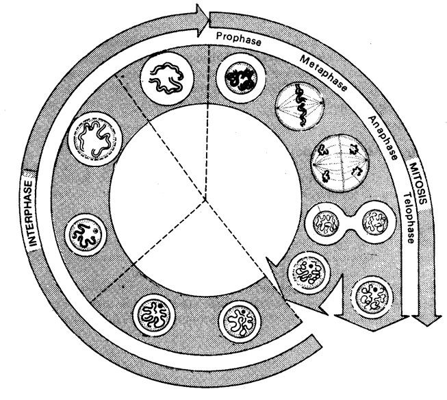
Active protein and
RNA synthesis
30
–
40
%
DNA replication
30
–50%
5
–10%
M
G
1
G
2
S
Growth
10
–20%
- Synthesis of ATP molecule and storage.
- Duplication of mitochondria, plastids and other cellular macromolecular complements.
- Damaged DNA repair occur.
(ii) M-phase/Dividing phase/Mitotic phase
- Nuclear division i.e. karyokinesis occurs in 4 phases – prophase, metaphase, anaphase and telophase. It takes 5-10% (shortest phase) time of whole division.
- Cytokinesis : Division of cytoplasm into 2 equal parts. In animal cell, it
takes place by cell furrow method and in Fig : Different stages of cell cycle (Mitotic cycle).
plant cells by cell plate method.
- Duration of cell cycle : It depends on the type of cell and external factors such as temperature, food and oxygen. Time period for G1, S, G2 and M-phase is species specific under specific environmental conditions.
e.g. 20 minutes for bacterial cell, 8-10 hours for intestional epithelial cell, and onion root tip cells may take 20 hours.
- Regulation of cell cycle : Stage of regulation of cell cycle is G1 phase during which a cell may follow one of the three options.
- It may start a new cycle, enter the S-phase and finally divide.
- It may be arrested at a specific point of G1 phase.
- It may stop division and enter G0 quiscent stage. But when conditions change, cell in G0 phase can resume the growth and reenter the G1 phase.
Types of cell division : It is of three types, Amitosis, Mitosis and Meiosis.
Important tips
- G0 – phase : The cells, which are not to divide further, do not proceed beyond the G1 phase and start undergoing differentiation into specific type. such cells are said to be in G0 phase.
- Generation time : Period between 2 successive generation (range 8 hr – 100 days).
- Mitogens : Chemicals which enhance or stimulate cell division e.g. lymphokinase (in man)
- Cell cycle duration : 20 minutes in bacteria , 20 hrs in root tip of onion, 2-3 hrs in yeast, 24 hrs in man.
- G0 phase : Cell only starts dividing when the period is favorable otherwise, it remain viable for months or years as such in G0 phase.
- During the mitosis of He-La cells, the longest period is gap I phase or G1.
- DNA replication occurs in S-phase.
- In a cell cycle the condensation of chromosome with visible centromere occurs during M-phase.
- Sequence in cell cycle is G SG M1, , 2, .
- M-phase is of shortest duration of cell cycle.
- In G2 , the damaged DNA is repaired.
- Histone protein and RNA synthesis occurs in S-phase.
- Duplication of chromosome occurs at S- phase.
Amitosis : (Gk amitos = without thread, osis = state) It is also called as direct cell division. It was discovered by Remak (1855) in RBC of chick embryo. In this division there is no Nucleus differentiation of chromosomes and spindle. The nuclear envelope does not degenerate.The nucleus elongates and constricts in the middle to form two daughter nuclei. This is followed by a centripetal constriction of the cytoplasm to form two daughter cells. It is primitive type of division occuring
in prokaryotes, protozoans, yeasts, foetal membrane of mammals, cartilage Plasma
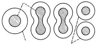
Daughter
membrane Cells of mammals, degenerating cells of the diseased tissues and in the old tissues.
Fig : Amitosis Mitosis : (Gk. Mitos = thread; osis = state)
- Definition : It is also called indirect cell division or somadtic cell division or equational division. In this, mature somatic cell divides in such a way that chromosomes number is kept constant in daughter cells equal to those in parent cell, so the daughter cells are quantitatively as well as qualitatively similar to the parental cell. So it is called equational division.
- Discovery : Mitosis was first observed by Strasburger (1875) and in animal cell by W.fleming (1879) term mitosis was given by Fleming (1882).
- Occurrence : Mitosis is the common method of cell division. It takes place in the somatic cells in the animals. Hence, it is also known as the somatic division. It occurs in the gonads also for the multiplication of undifferentiated germ cells. In plants mitosis occurs in the meristematic cells e.g. root apex and shoot apex.
- Duration : It ranges from 30 minutes to 3 hours time is species-specific but also depends upon type of tissues, temperature.
- Process of mitosis : Mitosis is completed in two steps
Karyokinesis : (Gk. karyon = nucleus; kinesis = movement) Division of nucleus. Term given by Schneider (1887).
Cytokinesis : (Gk –kitos = cell; kinesis = movement) Division of cytoplasm, Term given by Whitemann (1887).
Karyokinesis : It comprises four phases i.e. Prophase, Metaphase, Anaphase, Telophase.
(i) Prophase : It is largest phase of karyokinesis.
- Chromatin fibres thicken and shorter to form chromosomes which may overlap each other and appears
like a ball of wool. i.e. Spireme stage.
- Each chromosome divides longitudinally into 2 chromatids which remain attached to centromere.
- Nuclear membrane starts disintegrating except in dinoflagellates.
- Nucleolus starts disintegrating.
- Cells become viscous, refractive and oval in outline.
- Spindle formation begins.
- Cell cytoskeleton, golgi complex, ER, etc. disappear.
- In animal cells, centrioles move towards opposite sides.
- Lampbrush chromosomes can be studied well.
- Small globular structure (beaded) on the chromosome are called chromomeres.
(ii) Metaphase
- Chromosomes become maximally distinct i.e. size can be measured.
- A colourless, fibrous, bipolar spindle appears.
- Spindle is formed from centriole (in animal cells) or MTOC (microtubule organising centre) in plant cells successively called astral and anastral spindle.
- Spindle has 3 types of fibres.
- Continuous fibre (run from pole to pole).
- Discontinuous fibre (run between pole to centromeres).
- Interzonal fibre (run between 2 centromere).
(e) Spindle fibre are made up of 97% tubulin protein and 3% RNA.
No
Centrosome
Nucleus
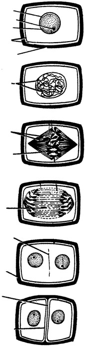
Spindle
fibres
Telophas
Metaphase
Metaphase
Prophase
Interphase
Nucleolus
Nuclear membrane
Plasma membrane
Cell wall
Chromo somes
Equator of spindle
Equatorial Plate
Cell plate
Cell wall
New cell wall
New plasma membrane
Divided cell
Fig : Various stages of mitosis
- Chromosomes move towards equatorial plane of spindles called congression and become arranged with their arms directed towards pole and centromere towards equator.
- Spindle fibres attach to kinetochores.
- Metaphase is the best stage for studying chromosome morphology.
(iii) Anaphase
- Centromere splits from the middle and two chromatids gets separated.
- Both the chromatids move towards opposite poles due to repulsive force called anaphasic movement.
- Anaphasic movement is brought about by the repolymerisation of continuous fibres and depolymerisation of chromosomal fibres.
- Different shape of chromosomes become evident during chromosome movement viz. metacentric acrocentric etc.
- Chromosomes takes V, J, I or L shapes.
- The centromere faces towards equator.
- The chromatids are moved towards the pole at a speed of 1 µm/minute. About 30 ATP molecules are used to move one chromosome from equator to pole.
(iv) Telophase
- Chromosomes reached on poles by the spindle fibers and form two groups.
- Chromosomes begin to uncoil and form chromatin net.
- The nuclear membrane and nucleolus reappear.
- Two daughter nuclei are formed.
- Golgi complex and ER etc., reform.
Furrow
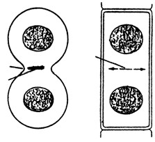
Cell
plate
Cytokinesis : It involves division of cytoplasm in animal cells, the Mid body cell membrane develops a constitution which deepens centripetally and is called cell furrow method.
In plant cells, cytokinesis occurs by cell plate formation. Fig : Furrowing Cell plate formation
(In animals) (In plants)
(6) Significance of mitosis
- It keeps the chromosome number constant and genetic stability in daughter cells, so the linear heredity of an organism is maintained. All the cells are with similar genetic constituents.
- It helps in growth and development of zygote into adult through embryo formation.
- It provides new cells for repair and regeneration of lost parts and healing of the wounds.
- It helps in asexual reproduction by fragmentation, budding, stem cutting, etc.
- It also restores the nucleo-plasmic ratio.
- Somatic variations when maintained by vegetative propagation can play important role in speciation.
(7) Types of Mitosis
- Anastral mitosis : It is found in plants in which spindle has no aster.
- Amphiastral mitosis : It is found in animals in which spindle has two asters, one at each pole of the spindle. Spindle is barrel-like.
- Intranuclear or Promitosis : In this nuclear membrane is not lost and spindle is formed inside the nuclear membrane e.g. Protozoans (Amoeba) and yeast. It is so as centriole is present within the nucleus.
- Extranuclear or Eumitosis : In this nuclear membrane is lost and spindle is formed outside nuclear membrane e.g. in plants and animals.
- Endomitosis : Chromosomes and their DNA duplicate but fail to separate which lead to polyploidy e.g.
in liver of man, both diploid (2N) and polyploid cells (4N) have been reported. It is also called endoduplication and endopolyploidy.
- Dinomitosis : In which nuclear envelope persists and microtubular spindle is not formed. During movement the chromosomes are attached with nuclear membrane. Important tips
- Pericentriolar cloud : A clear cytoplasmic area with no cell organelle between the centriole pair and astral rays.
- Root tips of onion are best material for studying mitosis.
- Kinetochore : A discoidal area on each chromatid and is the site of attachment of spindle fibres.
- In mitosis, plectonemic coiling takes place, in which sister chromatids are tightly coiled upon each other and are not easily separable.
Paranemic coiling found in meiosis.
- Chromosomal fibres are also called tractile fibres, while continuous fibres are also called interpolar fibres.
- Mitogens : The agents which stimulate cell division e.g., cytokinins, auxins, gibberllins, insulin, temperature, steroids.
- Mitotic poision : The agents which inhibit cell division.
- Azides and Cyanides : Inhibit prophase.
- Colchicine : Inhibits spindle formation at metaphase.
- Mustard gas : Agglutinates the chromosomes.
- Chalones : These were first reported by Laurence and Bullough (1960). They are peptides and glycoproteins secreated by extracellular fluid of healthy cells and inhibit cellular division.
- Karyochoriosis : A type of mitosis in fungi in which is intranuclear nucleus divides by furrow formation.
- C-mitosis : Colchicine induced mitosis.
- After undergoing certain divisions, cells die. This is called as “ Hayflick limit”.
- Actinomycin D and tetracyclin inhibit cell division.
- 7 mitotic divisions occur to form embryosac in angiosperms.
- Mitosis index is the ratio of dividing and non-dividing cells.
Meiosis : (Gk. meioum = to reduce, osis = state)
-
- Definition : It is a special type of division in which the chromosomes duplicate only once, but cell divides twice. So one parental cell produces 4 daughter cells; each having half the chromosome number and DNA amount than normal parental cell. So meiosis is also called reductional division.
- Discovery : It was first demonstrated by Van Benden (1883) but was described by Winiwarter (1900). Term “meiosis” was given by Farmer and Moore (1905).
- Occurrence : It is found in special types and at specific period. It is reported in diploid germ cells of sex organs (e.g. primary spermatocytes of testes to form male gametes called spermotozoa and primary oocytes to form female gametes called ova in animals) and in pollen mother cells (microsporocytes) of anther and megasporocyte of ovule of ovary of flowers in plant to form the haploid spores. The study of meiosis in plants can be done in young flower buds.
- Process of meiosis : Meiosis is completed in two steps, meiosis I and meiosis II
Meiosis I : In which the actual chromosome number is reduced to half. Therefore, meiosis I is also known as reductional division or heterotypic division. It results in the formation of two haploid cells from one diploid cell. It is divided into two parts, karyokinesis I and cytokinesis I.
Karyokinesis I : It involves division of nucleus. It is divided into four phases i.e. prophase, metaphase, anaphase, telophase.
Prophase I : It is of longest phase of karyokinesis of meiosis. It is again divisible into five subphases i.e.
leptotene, zygotene, pachytene, diplotene and diakinesis.
(i) Leptotene/Leptonema
- Chromosomes are long thread like with chromomeres on it.
- Volume of nucleus increases.
- Chromatin network has half chromosomes from male and half from female parent.
- Chromosome with similar structure are known as homologous chromosomes.
- Leptonemal chromosomes have a definite polarization and forms loops whose ends are attached to the nuclear envelope at points near the centrioles, contained within an aster. Such peculiar arrangement is termed as bouquet stage (in animals) and syndet knot (in plants).
- E.M. (electron microscope) reveals that chromosomes are composed of paired chromatids, a dense proteinaceous filament or axial core lies within the groove between the sister chromatids of each chromosome.
- Lampbrush chromosome found in oocyte of amphibians is seen in leptotene.
(ii) Zygotene/Zygonema
- Pairing or “synapsis” of homologous chromosomes takes place in this stage.
- Synapsis may be of following types.
- Procentric : Starting at the centromere.
- Proterminal : Starting at the end.
- Localised random : Starting at various points.
- Paired chromosomes are called bivalents, which by furthur molecular packing and spiralization becomes shorter and thicker.
- Pairing of homologous chromosomes in a zipper-fashion. Number of bivalents (paired homologous chromosomes) is half to total number of chromosomes in a diploid cell. Each bivalent is formed of one paternal and one maternal chromosome (i.e. one chromosome derived from each parent).
- Under EM, a filamentous ladder like nucleoproteinous complex, called synaptinemal. Complex between the homologous chromosomes which is discovered by “Moses” (1953).
(iii) Pachytene/Pachynema
- In the tetrad, two similar chromatids of the same chromosome are called sister chromatids and those of two homologous chromosomes are termed non-sister chromatids.
- Crossing over i.e. exchange of segments between non-sister chromatids of homologous chromosome occurs at this stage.
It takes place by breakage and reunion of chromatis segments. Breakage called nicking, is assisted by an enzyme endonuclease and reunion termed annealing is added by an enzyme ligase. Breakage and reunion hypothesis proposed by Darlington (1937).
- Chromatids of pachytene chromosome are attached with centromere.
- A tetrad consists of two sets of homologous chromosomes each with two chromatids. Each tetrad has four kinetochore (two sister and two homologous).
- A number of electron dense bodies about 100 nm in diameter are seen at irregular intervals within the centre of the synaptonemal complex, known as recombination nodules.
- DNA polymerase is responsible for the repair synthesis.
(iv) Diplotene/Diplonema
- At this stage the paired chromosomes begin to separate (desynapsis).
- Cross is formed at the place of crossing over between non-sister chromatids.
- Homologous chromosomes move apart they remain attached to one another at specific points called chiasmata.
- At least one chiasma is formed in each bivalent.
- Chromosomes are attached only at the place of chiasmata.
- Chromatin bridges are formed in place of synaptonemal complex on chiasmata.
- This stage remains as such for long time.
(v) Diakinesis
- Chiasmata moves towards the ends of chromosomes. This is called terminalization.
- Chromatids remain attached at the place of chiasma only.
- Nuclear membrane and nucleolus degenerates.
- Chromosome recondense and tetrad moves to the metaphase plate.
- Formation of spindle.
- Bivalents are irregularly and freely scattered in the nucleocytoplasmic matrix.
When the diakinesis of prophase-I is completed than cell enters into the metaphase-I.
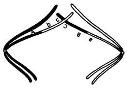
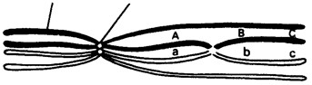
Chromatid
Centromere
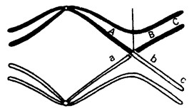
Chiasma
Fig : Showing crossing over during meiosis
Chromosome
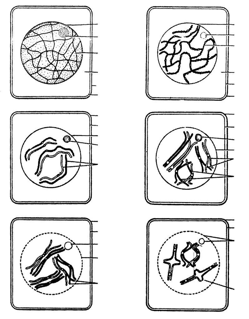
Nuclear
membrane
Nuclcolus
Nuclcolus
Nuclear
membrane
Chromosome
network
Cytoplasm
Cell wall
Cell membrane
Cell wall
Cell wall
Cell wall
Cell membrane
Nuclear membrane
Nuclcolus
Pair of
homologous
chromosomes
showing synspsis
Pair of sister
chromatids
Non-
Cell wall
Cell membrane
Nuclcolus
Nuclear membrane
disspearing
Homologous
chromosomes
showing chlasma
A
B
C
D
E
F
showing network
chromomeres
Cytoplasm
Cell membrane
Cell membrane
Nuclcolus
Nuclear membrane
soster chromatids of
homologous chromosomes
showing crossing over
Cell wall
Cell membrane
Disappearing
nucleolus and
nuclear membrane
Homologous
chromosomes separating from
one another terminalleation
Fig : A-F Meiosis : Prophase I. A. Cell before entering leptotene, B. Leptotene, C. Zygotene, D. Pachytene, E. Diplotene, F. Diakinesis.
Metaphase I : It involves;
- Chromosome come on the equator.
- Due to repulsive force the chromosome segment get exchanged at the chiasmata.
- Bivalents arrange themselves in two parallel equatorial or metaphase plates. Each equatorial plate has one genome.
- Centromeres of homologous chromosomes lie equisdistant from equator and are directed towards the poles while arms generally lie horizontally on the equator.
- Each homologous chromosome has two kinetochores and both the kinetochores of a chromosome are joined to the chromosomal or tractile fibre of same side.
Anaphase-I
- It involves separartion of homologous chromosomes which start moving opposite poles so each tetrad is divided into two daughter dyads. So anaphase-I involves the reduction of chromosome number, this is called disjunction.
- The shape of separating chromosomes may be rod or J or V-shape depending upon the position of centromere.
- Segregation of mendalian factors or independent asortment of chromosomes take place. In which the paternal and maternal chromosomes of each homologous pair segregate during anaphase-I which introduces genetic variability.
Telophase-I
- Two daughter nuclei are formed but the chromosome number is half than the chromosome number of mother cell.
- Nuclear membrane reappears.
- After telophase I cytokinesis may or may not occur.
- At the end of Meiosis I either two daughter cells will be formed or a cell may have two daughter nuclei.
- Meiosis I is also termed as reduction division.
- After meiosis I, the cells in animals are reformed as secondary spermatocytes or secondary oocytes; with haploid number of chromosomes but diploid amount of DNA.
(vi) Chromosomes undergo decondensation by hydration and despiralization and change into long and thread like chromation fibres.
Interphase : Generally there is no interphase between meiosis-I and meiosis-II. A brief interphase called interkinesis, or intermeiotic interphase. There is no replication chromosomes, during this interphase.
Cytokinesis-I : It may or may not be present. When present, it occurs by cell-furrow formation in animal cells and cell plate formation in plant cells.
Significance of meiosis-I :
- It separates the homologous chromosomes to reduce the chromosome number to the haploid state, a necessity for sexual reproduction.
- It introduces variation by forming new gene combinations through crossing over and randon assortment of paternal and maternal chromosomes.
- It may at times cause chromosomal mutation by abnormal disjunction.
- It induces the cells to produce gametes for sexual reproduction or spores for asexual reproduction.
Meiosis-II : It is also called equational or homotypical division because the number of chromosomes remains same as after meiosis-I. It is of shorter duration than even typical mitotic division. It is also divisible into two parts, Karyokinesis-II and Cytokinesis-II.
Karyokinesis-II : It involves the separation of two chromatids of each chromosome and their movement to separate cells. It is divided in four phases i.e., Prophase-II, Metaphase-II. Anaphase-II and Telophase-II.
Almost all the changes of Karyokinesis-II resembles to mitosis which involves.
- It starts just after end of telophase I.
- Each daughter cell (nucleus) undergoes mitotic division.
- It is exactly similar to mitosis.
- At the end of process, cytokinesis takes place.
- Four daughter cells are formed after completion.
- The sister kinetochores of one chromosome are separated.
- The four daughter cells receive one chromatid each of the tetravalent.
- Centromere divide at anaphase II.
- Spindle fibres contract at prophase II.
Cytokinesis-II : It is always present and occurs by cell furrow formation in animal cell and cell plate formation in plant cell.
So by meiosis, a diploid parental cell divides twice forming four haploid gametes or sex cells, each having half the DNA amount than that of the parental cell and one-fourth of DNA present in the cell at the time of beginning of meiosis.
(5) Significance of meiosis
- Constancy of chromosome number in successive generation is brought by process.
- Chromosome number becomes half during meiosis.
- It helps in introducing variations and mutation.
- It brings about gamete formation.
- It maintains the amount of genetic informative material.
- Sexual reproduction includes one meiosis and fusion.
- The four daughter cells will have different types of chromatids.
(6) Why the necessity of meiosis-II : The basic aim of meiosis is to reduce the number of chromosomes to half. The chromosomes that separate in the anaphase of meiosis-I are still double. Each consist of two chromatids and has 2n amount of DNA. Thus reduction of DNA content does not occur in meiosis-I. Truely haploid nuclei in terms of DNA contents as well as chromosome number are formed in meiosis-II. When the chromatids of each chromosome are separated into different nuclei. Thus meiosis-II is necessary.
Difference between Mitosis and Meiosis
| S.No. | Characters | Mitosis | Meiosis |
| I. General | |||
| (1) | Site of occurrence | Somatic cells and during the multiplicative phase of gametogenesis in germ cells. | Reproductive germ cells of gonads. |
| (2) | Period of occurrence | Throughout life. | During sexual reproduction. |
| (3) | Nature of cells | Haploid or diploid. | Always diploid. |
| (4) | Number of divisions | Parental cell divides once. | Parent cell divides twice. |
| (5) | Number of daughter cells | Two. | Four. |
| (6) | Nature of daughter cells | Genetically similar to parental cell. Amount of DNA and chromosome number is same as in parental cell. | Genetically different from parental cell. Amount of DNA and chromosome number is half to that of parent cell. |
| II. Prophase | |||
| (7) | Duration | Shorter (of a few hours) and simple. | Prophase-I is very long (may be in days or months or years) and complex. |
| (8) | Subphases | Formed of 3 subphases : early-prophase, mid-prophase and late-prophase. | Prophase-I is formed of 5 subphases: leptotene, zygotene, pachytene, diplotene and diakinesis. |
| (9) | Bouquet stage | Absent. | Present in leptotene stage. |
| (10) | Synapsis | Absent. | Pairing of homologous chromosomes in zygotene stage. |
| (11) | Chiasma formation and crossing over. | Absent. | Occurs during pachytene stage of prophase-I. |
| (12) | Disappearance of nucleolus and nuclear membrane | Comparatively in earlier part. | Comparatively in later part of prophase-I. |
| (13) | Nature of coiling | Plectonemic. | Paranemic. |
| III. Metaphase | |||
| (14) | Metaphase plates | Only one equatorial plate | Two plates in metaphase-I but one plate in metaphase-II. |
| (15) | Position of centromeres | Lie at the equator. Arms are generally directed towards the poles. | Lie equidistant from equator and towards poles in metaphase-I while lie at the equator in metaphase-II. |
| (16) | Number of chromosomal fibres | Two chromosomal fibre join at centromere. | Single in metaphase-I while two in metaphase-II. |
| IV. Anaphase | |||
| (17) | Nature of separating chromosomes | Daughter chromosomes (chromatids with independent centromeres) separate. | Homologous chromosomes separete in anaphase-I while chromatids separate in anaphase in anaphase-II. |
| (18) | Splitting of centromeres and development of inter-zonal fibres | Occurs in anaphase. | No splitting of centromeres. Inter-zonal fibres are developed in metaphase-I. |
| V. Telophase | |||
| (19) | Occurrence | Always occurs | Telophase-I may be absent but telophase-II is always present. |
| VI. Cytokinesis | |||
| (20) | Occurrence | Always occurs | Cytokinesis-I may be absent but cytokinesis-II is always present. |
| (21) | Nature of daughter cells | 2N amount of DNA than 4N amount of
DNA in parental cell. |
1 N amount of DNA than 4 N amount of
DNA in parental cell. |
| (22) | Fate of daughter cells | Divide again after interphase. | Do not divide and act as gametes. |
|
VII. Significance |
|||
| (23) | Functions | Helps in growth, healing, repair and multiplication of somatic cells.
Occurs in both asexually and sexually reproducing organisms. |
Produces gametes which help in sexual reproduction. |
| (24) | Variations | Variations are not produced as it keeps quality and quantity of genes same. | Produces variations due to crossing over and chance arrangement of bivalents at metaphase-I. |
| (25) | In evolution | No role in evolution. | It plays an important role in speciation and evolution. |
(7) Types of meiosis : On the basis of time and place, meiosis is of three types
- Gametic/Terminal meiosis : In many protozoans, all animals and some lower plants, meiosis takes place before fertilization during the formation of gametes. Such a meiosis is described as gametic or terminal.
This type of life cycle with diploid adult and gametic meiosis is known as the diplontic cycle.
- Zygotic or Initial Meiosis : In fungi, certain protozoan groups, and some algae fertilization is immediately followed by meiosis in the zygote, and the resulting adult organisms are haploid. Such a meiosis is said to be zygotic or initial. This type of life cycle with haploid adult and zygotic meiosis is termed the haplontic cycle.
(iii) Sporogenetic Meiosis
- Diploid sporocytes or spore mother cells of sporophytic plant, undergo meiosis to form the haploid spores in the sporangia.
- Haploid spore germinates to form haploid gametophyte which produces the haploid gametes by mitosis.
- Haploid gametes fuse to form diploid zygote which develops into diploid sporophyte by mitotic divisions.
e.g. In higher plants like pteridophytes, gymnosperms and angiosperms.
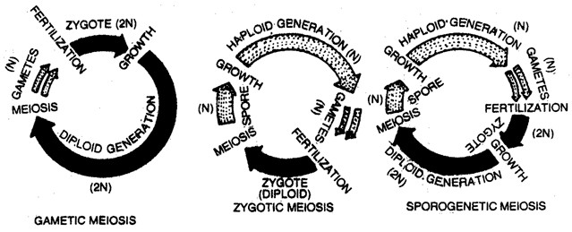
Fig : Three types of Meiosis
Important tips
- Brachymeiosis : Failure of meiosis-II. It is characteristic feature of fungi.
- Meiosis-II is not mitosis as it occurs haploid number of chromosomes and chromatids formed may not be similar to each other.
- Restitution nucleus : A colchicine treated cell has the nucleus with double sets of chromosomes.
- In cyperus, one meiosis produce only one pollen instead of four so that meiotic division required to produce fruits will be = number of fruits × 2.
- Chiasmata first observed by Janseens (1909).
- When sister chromatids are loosely arranged and are easily separate. It is found in meiotic chromosomes.
- Mitosis ends in 1- 2 hours while meiosis may take 24 hrs to few years.
- Neuron cells are always in interphase.
- When the chromosome duplicates but karyokinesis does not take place the number of chromosome per cell will increases, it is called endomitosis or endoduplication.
- Process of inducing mitosis into a cell – mitogenesis.
- To study mitosis root tips are fixed in 1: 3 acetic acid and methanol.
- Colchicine inhibits spindle formation and enhance duplication in number of chromosomes.
- At the time of cell division electrostatic force is responsible for terminalization.
- Mitotic crossing over takes place in parasexual cycle.
| Introduction. |
There are millions of organisms – plants, animals, bacteria and viruses. Each one is different from the other in one way or the other. About more than one million of species of animals and more than half a million species of plants have been studied, described and provided names for identification. Thousands are still unknown and are yet to be identified and described. It is practically impossible to study each and every individual. Also, it is difficult to remember their names, characters and uses. However, biologists have devised techniques for identification, naming and grouping of various organisms.
The art of identifying distinctions among organisms and placing them into groups that reflect their most significant features and relationship is called biological classification. Scientists who study and contribute to the classification of organisms are known as systematists or taxonomists, and their subject is called systematics (Gk. Systema = order of sequence) or taxonomy (Gk. Taxis = arrangement; nomos = law).
History of classification : References of classification of organisms are available in Upanishads and Vedas. Our Vedic literature recorded about 740 plants and 250 animals. Few other significant contributions in the field of classification are :
-
- Chandyogya upanishad : In this, an attempt has been made to classify the animals.
- Susruta samhita : It classifies all ‘substances’ into sthavara (imbobile) e.g. plants and jangama (mobile) e.g. animals .
- Parasara : Here, angiosperms were classified into dvimatruka (dicotyledons) and ekamatruka (monocotyledons). He was even able to find that dicotyledons bear jalika parana (reticulate veined leaves and monocotyledons bear maun laparna parallel veined leaves).
- Hippocrates, and Aristotle : They classified animals into four major groups like insects, birds, fishes and whales.
| Types of system of classification. |
Different systems of classification proposed from time to time have been divided into three basic categories viz., artificial systems, natural systems and phylogenetic system (However, Redford, (1986), included mechanical systems as a fourth category).
-
- Artificial system of classifications : These systems are more or less arbitrary as the plants are classified merely on the basis of gross morophology, habit and their importance to man. The main advocates of artificial system of classifications were :
- Theophrastus : Father of botany. Theophrastus was a disciple of Plato and later Aristotle. In his book De Historia plantarum, he classified about 500 kinds of plants into four major group; trees, shrubs, subshrubs and herbs.
- Caius Plinius Secundus : He described the biological, medicinal and agricultural aspects of plants in 37 volumes of Natural History. He used the term ‘Stamen’ for the first time.
- Pedanios Dioscorides : He described about 600 plants of medicinal importance in his Materia Medica.
- Charaka : Indian Scholar. He classified plants of medicinal importance in his Charaka Samhita.
- Andrea Caesalpino : He described 1520 species in 16 volumes of De Plantis libri grouped as herbs and trees. He further classified plants based on fruit and seed characters.
- John Ray : He was a British botanist who published three volumes of his work Historia Generalish Plantarum consisting of improved classification originally proposed by him in Methodus Plantarum Noven. He was the first to divided the groups herbs, shrubs and trees into Dicots and Monocots on the basis of the presence of two or one cotyledons respecitvely. He coined the term species.
- Carolus Linnaeus : Father of taxonomy. A swedish botanist, who published an artificial system of classification based exclusively on floral characters. Linnaeus published several manuscripts including Hortus cliffortianus and Genera plantarum (1737). In his Genera plantarum he listed all the plant genera known to him. He published his best known Species plantarum in 1753. In this book he listed and described all species of plants known to him. He established binomial nomenclature.
- Natural System of Classifications : These systems of classification are based not only on the characters of reproductive organs and structural morphology but used as many taxonomic characters or traits as possible to classify the plants. The advocates of natural systems of classification are listed below :
- Michel Adanson : A French botanist, who classified plants and animals using as many characters as possible and proposed a natural system of classification.
- A.L. de Jussieu : Classified plants based on natural characters. In his system of classification he grouped the plants resembling each other in a set of characters.
- A.P. de Candolle : He grouped all alike plants together and published a new classification of plants in his book Theorie elementaire de la botanique (1813).
- George Bentham and Joseph Dalton Hooker : These two English botanists classified plants based on original studies of specimens. They published their well known scheme of classification in Genera plantarum (1862– 83). This system of classification is still regarded as the best classification, especially from the practical point of view.
Bentham and Hooker’s classification (Broad outline)
Plant Kingdom
Cryptogams
Non-flowering plants)
(
Phanerogams
(
Seed bearing plants)
Dicotyledonae
Seed having two cotyledons)
(
Monocotyledonae
Seed having one cotyledons)
(
Class
Gymnospermae
Polypetalae
Petals free)
(
Sub-Class
Gamopetalae
Petals fused)
(
Monochlamydeae
(
Petals absent)
Series
Inferae
(
Ovary inferior)
e.g.
, Compositae
Bicarpellatae
(
Ovary superior, Bicarpellary)
e.g.
, Solanaceae, Labiatae
Heteromerae
Thalamiflorae
Petals and stamens hypogynous)
(
e.g.
, Ranunculaceae
Brassicaceae, Malvaceae
Disciflorae
Petals and stamens
(
hypogynous, a nectariferous
disc at the base of the ovary)
e.g.
, Rutaceae
Calyciflorae
(
Petals and stamens
perigynous/epigynous)
e.g.
, Leguminosae, Cucurbitaceae
Series
Microspermae
Epigynae
(
Ovary
Coronarieae
Perianth petaloid, ovary
Calycinae
Nudiflorae
Apocarpae
Free
(
Glumaceae
Perianth small,
(
inferior) superior e.g., Liliaceae carpels) scale-lke or chaffy
e.g., Gramineae)
Merits and Demerits of Bentham and Hooker’s system of classification Merits
This system is regarded as most convenient and suitable for practical utility and is followed by most of the herbaria due to following reasons :
- Every genus and species was studied from actual specimens available in British and continental herbaria.
- It is the first great natural system of classification. This is very useful for practical purposes.
- Gymnosperms have been considered as third taxon and kept between dicots and monocots.
- In monocots, stress has been given to relative position of ovary and perianth characteristics.
- Great emphasis has been given to free or fused conditions of petals distinguishing dicots into three subclasses Polypetalae, Gamopetalae and Monochlamydeae.
Demerits : There are some demerits in the classification of Bentham and Hooker. Some of them are :
- The greatest demerit of this system is the retention in the group monochlamydeae, a number of orders which represent affinities with those in which biseriate perianth is the rule.
- Placing gymnosperms between dicots and monocots.
- Here monochlamydeae is considered as most highly evolved and polypetalae as primitive among dicots.
- Cucurbitaceae family with fused petals is placed in Polypetalae.
- Liliaceae is separated from Iridaceae and Amaryllidaceae merely on the character of ovary, without keeping in mind other similarities.
Differences between Natural and Artificial classification
| No. | Characters | Natural classification | Artificial classification |
| 1. | Number of characters | Almost all the characters are considered. | Only few characters are considered. |
| 2. | Hereditary constitution | Members are mostly alike in hereditary pattern of different groups. | Members of different groups are usually not similar in hereditary pattern. |
| 3. | Flexible | May change with advancement in knowledge. | Stable classification. |
| 4. | Phylogeny | Closely related phylogenetically. | Not related phylogenetically. |
| 5. | Information | Provides plenty of useful information. | Provides only limited information. |
| 6. | Recent advances | Recent useful research can be easily incorporated. | Cannot incorporate new work. |
| 7. | Convenience | Identification of plants easy. | Difficult. |
| 8. | Little known plants | Got the place at definite place. | Not certain about position and identification. |
(3) Phylogenetic system of classifications : These systems of classifications are mainly the rearrangements of natural systems using as many taxonomic characters as possible in addition to the phylogenetic (evolutionary) informations. Some important phylogenetic systems of classifications were proposed by –
- A.W. Eichler : A German botanist who proposed phylogenetic system of classification and published in the third edition of Syllabus der vorlesungen (1883).
- Adolph Engler and Karl Prantl : These two german botanists classified plant kingdom on the basis of their evolutionary sequences. They started with simplest flowering plants and ended with plants of complex floral structures.
- C.E. Bessey : He classified flowering plants on the basis of their evolutionary relationships.
- John Hutchinson : A British botanist published his phylogenetic system of classification in ‘The Families of Flowering Plants‘.
- Armen Takhtajan : A Russian botanist who published his system of classification in Botanical Review.
- Arthur Cronquist : Published his classification in ‘An Integrated System of Classification of Flowering Plants‘.
Differences between Natural and Phylogenetic classification
| No. | Characters | Natural classification | Phylogenetic classification |
| 1. | Number of characters | Based on several constant characters. | Along with several constant characters, evolutionary sequences are also considered. |
| 2. | Evolutionary sequences | Not considered. Classification based basically on common characters. | Evolutionary sequence, natural affinities and relationship taken into account. |
| 3. | Practical utility | Used adequately as an aid for easy identification. | Phylogenetic and adopted by many countries. |
| Plant nomenclature. |
Plant nomenclature may be defined as the system of naming plant. Almost all plants (and animals too) are known by different common names in different parts of the world. Even within the same country people of different states and regions use different common names. Iphomoea batatas, for example, is called sweet potato in English, Shakarkandi in Hindi, Meetha alu in Assamies and Bengali, Kundmul in Telagu, Ratalu in Marathi and Jenasu in Kannad. The common names are thus quite confusion. This necessiated the need of giving scientific names so that scientists of different parts of the world could understand each other work. The earliest scientific names were polynomial, i.e., they were composed of many words (which gave the characteristics of plants), e.g., Sida acuta (a member of Malvaceae) was named as Chrysophylum foliis, ovalis superne glabris parallel striatis subtus, tomentosonitidis. Such long names were difficult to remember. Hence, to make it easier binomial system of nomenclature was introduced.
(1) Binomial system of nomenclature : The credit of giving binomial system of nomenclature goes to
Swedish naturalist, Carolus Linnaeus. He employed this system in his book Species Plantarum, published in 1753. According to this system the name of a plant or animal is composed of two Latin (or Latinised) words, e.g., potato is Solanum tuberosum. The first word (i.e., Solanum) indicates the name of the genus (called generic name) and the second word (i.e., tuberosum) denotes the name of the species (called specific name). The generic name always begins with a capital letter and the specific name with a small letter and printed in italics.
The generic and specific names always have some meaning. They are based on some special characters of the plant, on the name of any scientist or on some legend.
All plants having general similarity and relations are given a common generic name, e.g., potato, brinjal, black nightshade (makoi) have been placed in the genus Solanum. However, their specific names distinguish them from each other – potato is Solanum tuberosum, brinjal is S. melongena and black nightshade is S. nigrum.
Usually the name of the author, who names a plant, is also written in full or in abbreviated form after the specific name. Thus, in case of Mangifera indica L., the L. stands for Linnaeus and in Lychnis alba Mill., the Mill. stands for Miller.
Sometimes a single species is described under different names by different authors. These name are called synonyms. In such cases, the name under which the species is first described, is considered to be valid.
- Trinomial nomenclature : Certain species are divisible into smaller units, called varieties, on the basis of finer differences. The name of the variety is written after the specific name. Thus, the name may become trinomial or three word name. e.g., Homo sapiens europeus is the name of the man of European race. Trinomial nomenclature is simply an extension of the Linnaean system.
- Code of biological nomenclature : Anyone can study, describe, identify and give a name to an organism provided certain universal rules are followed. These rules are framed and standardised by International Code of Botanical Nomenclature (ICBN) and International Code of Zoological Nomenclature (ICZN). The codes help in avoiding errors, duplication, confusion and ambiguity in scientific names. The codes are established and improved upon at International Botanical and Zoological Congress held from time to time. The names of bacteria and viruses are decided by International Code of Bacteriological Nomenclature (ICBN) and International Code of Viral Nomenclature (ICVN). Similarly, there is a separate International Code of Nomenclature for Cultivated Plants (ICNCP).
| Explanation of terminology (Units). |
- Taxon : The term taxon is used to represent any unit of classification. The unit (i.e., taxon) many be large (e.g., Plant Kingdom) or small (e.g., Algae, Fungi, or a single species).
- Category : Various sub-divisions of plants kingdom such as division, class, order, family, etc., are referred to as categories. In the hierarchy of categories kingdom is the highest and species is the lowest category. The following is hierarchial series :
- Kingdom : It is the highest category in biological classification. All plants are include in plant kingdom.
- Division : It is a major group in the Linnean hierarchy used in the classification of plants (equivalent to phylum in animal classification). It is a taxonomic category between kingdom and class. The subcategory of division is subdivision. The suffix of division is – ophyta.
- Class : A division is divided into classes. It is a taxonomic category between the division and order. Its suffix is – ae. The subcategories of class are subclass and series. In the class contain organism least similar to one another.
- Order : A class includes one or more orders. It is a taxonomic category between the class and family. Its suffix is – ales. The subcategory of order is suborder.
- Family : An order is divided into one or more families. It is the taxonomic category between the order and the genus. Its suffix is – aceae. The subcategories of family are subfamily, tribe and subtribe.
- Genus : The plural of genus is genera. A family includes one or more genera. The generic name is important and printed in Italics (If hand written, it is underlined). The subcategories of genus are subgenus, section and subsection.
- Species : It is the smallest rank of taxonomic classification. The first letter of the species is denoted with small letter. The species is printed in Italics (It is underlined if hand written). A genus may include one or more species. The subcategories of species are subspecies, varieties, subvarieties, form and subform.
(3) New systematics or Biosystematics : The term new systematics was proposed by Sir Julian Huxley in
1940. In the new systematics, the species are considered related to one another, mutable and the work of gradual modification. This is in confirmity with the facts of evolution.
Forms of new systematics : There are several forms of new systematics – (i) Morphotaxonomy : It is based on the structural features of the organisms.
- Cytotaxonomy : It is based on the somatic chromosomes of organisms.
- Biochemical taxonomy or Chemotaxonomy : It is based on the protein and serum analyses and on the chemical constituents of the organisms.
- Numerical taxonomy : It involves quantitative assessment of similarities and differences in order to make objective assessments. Characters of organisms are given equal weight and the relationships of the organisms are numerically determined, usually with the aid of a computer.
- Experimental taxonomy : It is based on the genetic relationship determined with the help of experiments.
Important Tips
- The arrangement of organism in to groups termed as classification.
- Forming the rules for classification known as Taxonomy.
- De candolle : Coind the term taxomy in 1913.
- Floral characters are used as basis of classification and for identifying new species because floral characters are conservative when compared with vegetative characters.
- In Bentham and Hooker’s classification Dicotyledons have been kept before Monocotyledons. Seeds plants have been divided into Dicots, Gymnospermae and Monocots.
- Among the vegetative characters, venation in leaf in one of highly acceptable characters for classification of angiosperms.
- In Engler and Prantl’s system Monocotyledons have been kept before Dicotyledons.
- In Bentham and Hooker’s classification 202 families have been identified.
- Bentham and Hooker’s classification is a natural system of classification and very helpful for practical purposes.
- For declaration of new species, floral characters of new species should be used.
- In Bentham and Hooker’s system of classification, evolutionary criteria have not been followed hence not phylogenetic.
- Hooker complied first complete flora of India and wrote the book ‘Flora of British India’.
- Bentham and Hooker : Gave the first natural classification of plants.
- Engler and Prantl : Gave the first phylogenetic classification of plants.
- Engler and Prantl’s and Hutchinson’s system of classification are phylogenetic.
- 411 families have been recognised in Hutchinson’s system of classification and 280 families have been identify in Engler and Prantl’s system of classification.
- Father of Botany : Theophrastus, a Greek philosopher, produced the first book on botany, Historia Plantarum, student of Aristotle.
- After the work of Linnaeus, another significant publication was that of Augustin de Candolle in theory elementaire de la botanique.
- The correct sequence of taxa in Linnaean hierarchy is species → genus → family → order→ class.
- A system for naming the organisms called nomenclature.
- Bauhin (1623) proposed the binary system of nomenclature which was elaborated by Linnaeus (1753) in to binomial system.
- An international code of Botanical nomenclature (IBCN) come into existence in 1930.
- International code for nomenclature is divided into three parts i.e. (i) Principles (ii) Rules and recommendations (iii) Provisions.
- Priority : Nomenclature of taxonomic groups is based upon priority of publication.
- Monotypic Genus : A genus having only one species, e.g., Home.
- Polytypic Genus : A genus containing more than one species, e.g., Panthera, Solanum.
- John Ray : An English naturalist, 1627–1705, introduced the term species. It is a basic unit of classification.
- J.K. Maheswari described the plants of India in ‘Flora of Delhi’.
- Phylogeny was introduced by Homock but concept was introduced by Heackel.
- Phylogenetic classification reflect the evolutionary relationships of organisms.
- Type Specimen : Original specimen is called holotype; duplicate of holotype is termed isotype; additional one is known as paratype; and a new one when the original is lost is referred to as neotype.
| Modern system of classification. |
-
- Two kingdom system of classification : This system of classification is the oldest it was suggested by
Carolus Linnaeus in 1758. He divided the living word (organism) in to two kingdoms, Plantae (for all plants like tree, shrubs, climbers, creepers, moss and floating green algae) and Animalia (For animals).
-
- Three kingdom system of classification : Ernst Haeckel, a German biologist and philosopher, suggested a third kingdom protista in 1866 for –
- Unicellular organisms such as bacteria, protozoans and acellular algae.
- Multicellular organisms without tissue such as algae and fungi.
- Four kingdom system of classification : It was proposed by Copeland in 1956. The two additional kingdoms were Monera for the bacteria and blue green algae and Protista for protozoans, algae and fungi.
- Five kingdom system of classification : R.H. Whittaker, an American ecologist. He proposed five kingdom system of classification in 1969.
This system replaced the old, two-kingdom grouping of living organisms. As already discussed, a division of living world merely into plant and animal kingdoms is too simple. It does not take into account the gradual evolution of distinct plant and animal groups and it allows no place for those primitive organisms that even now are neither plants nor animals nor that are both. In this classification eukaryotes were assingned to only four of the five kingdom.
Five-kingdom classification is based on the following four criteria :
-
- Complexity of cell structure.
- Complexity of organism’s body.
- Mode of obtaining nutrition.
- Phylogenetic relationship.
The five kingdom are : Monera, Protista, Fungi, Plantae and Animalia.
| Kingdom Monera (The prokaryotes). |
Monera (Monos – single) includes prokaryotes and shows the following characters :
-
- They are typically unicellular organisms (but one group is mycelial).
- They lack nuclear membranes.
- Ribosomes and simple chromatophores are the only subcellular organelles in the cytoplasm. The ribosomes are 70 S. Mitochondria, plastids, Golgi apparateus, lysosomes, endoplasmic reticulum, centrosome, etc., are lacking.
- The predominant mode of nutrition is absorptive but some groups are photosynthetic or chemosynthetic.
- Reproduction is primarily asexual by fission or budding.
- Protosexual phenomenon also occurs.
- The organisms are non-motile or move by beating of simple flagella or by gliding.
- Flagella, if present, are composed of many intertwined chains of a protein flagellin. They are not enclosed by any membrane and grow at the tip.
- Moneran cells are microscopic (1 to few microns in length).
- Most organisms bear a rigid cell wall.
- The kingdom Monera includes true bacteria, mycoplasmas, rickettsias, actinomycetes (ray fungi) etc. Microbiologists also include blue green algae (i.e., Cyanobacteria) under the group bacteria because of the presence of prokaryotic cell structure. Studies have established that the members of archaebacteria group are most primitive and have separated from eubacteria group very early in the process of evolution. Furthermore, these studies have also concluded that the archaebacteria and eubacteria possibly originated from a more ancient form of life called Progenote.
- Nutrition : They show both autotrophic and heterotrophic modes of nutrition.
(i) Autotrophs : These are able to form their own food by one of the following methods.
-
- Photoautotrophs : They prepare their own food by reducing CO2 using light energy.
- Chemoautotrophs : They form their food by energy derived from chemical reaction.
(ii) Heterotrophs : A few live in symbiosis while others form association of commensalism. Saprophytes also called ‘saprobes’ cause decay, fermentation or putrefaction of dead organic matter. Some bacteria are facultative sasprophyte (= facultative parasites). In the process of fermentation there is anaerobic break-down of carbohydrates into CO2, alcohol and some energy. Putrefaction or decay is anaerobic break-down of proteins accompanied by foul smell due to evil smelling gases produced in the process.
The saprobes produce enzymes which convert non-diffusible food substrates (carbohydrate fats, proteins, etc.) into simpler diffusible form which diffuses into the cytoplasm and is assimilated, i.e., converted into body cytoplasm or stored as reserve food.
Still others live on other living organisms (animals, plants or man) in the form of parasites directly absorb their food from the body of host. Some of the parasites are non-pathogenic i.e., cause no ill-effect or disease in the host, while some are pathogenic causing diseases in the host.
| (d) Major ecological role | Producer Autotrophy | Decomposer
Heterotrophy absorption |
Consumer
Heterotrophy ingestion |
Kingdom
fungi
Kingdom
Animalia
Kingdom
Plantae
Direction of evolution
(
Multicellular)
Simple
(
uncellular)
Eukaryotes
Kingdom
protista
Kingdom
monera
Prokaryotes
Autotrophy
chemosynthesis
photosynthesis
Fig : Probable phylogenetic relationships among the kingdoms
(c) Mode of nutrition
(b) Complexity
of organism
(a) Complexity of cell
(13) Reproduction : It is primarily asexual by binary fission or budding. Mitotic apparatus is not formed during cell division. Distribution of replicated DNA into daughter cells is assisted by cell membrane. Exchange of genetic material between two bacterial cells is known to occur but no gametes are formed. Sterols, the precursor molecules of sex hormones, have been reported from certain prokaryotes. Many bacteria form resistant spores.
| Kingdom Protista (Unicellular eukaryotes). |
Protista (Protistos = Primary) includes unicelluar eukaryotes and show the following characters :
-
- Protists include solitary unicellular or colonial unicellular eukaryotic organisms which not form tissues.
- Simple multinucleate organisms or stages of life cycles occur in a number of groups.
- The organisms possess nuclear membranes and mitochondria.
- In many forms, plastids, (9+2 strand) flagella and other organelles are present.
- The nutritive modes of these organisms include photosynthesis, absorption, ingestion and combination of these.
- Some protists possess contractile vacuole for regulation of their water content.
- Their reproductive cycles typically include both asexual divisions of haploid forms and true sexual processes with karyogamy and meiosis.
- The organisms move by flagella or by other means or are non-motile.
- Nutrition : It is may be photosynthetic, holotrophic, saprotrophic and parasitic. Some have mixotrophic nutrition (holotrophic + saprobic). Chemosynthetic nutrition is lacking. Certain protozoans decompose organic matter, such as cellulose, in the gut of termites and woodroaches. They live as symbionts. The photosynthetic, floating protists are collectively called phytoplankton. They usually have a cell wall. The free-floating, holozoic protozoans are collectively termed zooplankton. They lack cell wall to allow ingestion of particulate food.
- Reproduction : It is occurs by both asexual and sexual methods :
(i) Asexual reproduction : It is the most common method of reproduction in protists in which the genetic constitutions of young ones remains the same as that of the parent. Under favourable environmental conditions, they reproduce asexually several times a day resulting in population explosions. The major types of asexual reproductions are as follows :
-
- Binary fission : The parent cell divides into two approximately equal daughter cells either transversely (e.g., Paramecium), longitudinally (e.g., Euglena) or axially (e.g., Amoeba) by mitosis.
- Multiple fission : Division of parent cell into a number of daughter cells is called multiple fission. It occurs in Amoeba.
- Plasmotomy : Fission of multinucleate protist into two or more multinucleate offsprings by the division of cytoplasm without nuclear division is called plasmotomy. It occurs in Opalina.
- Budding : In this type of asexual reproduction, a small bud is formed from the parent body which separates and develops into new individual. e.g., Paracineta, Arcella, etc.
- Spore formation : Sessile or stalked sporangia containing spores are formed in slime moulds. They liberate the spores which can withstand a prolonged period of desiccation. On germination, each spore gives rise to new individual. e.g., Slime moulds.
(ii) Sexual reproduction : Sexual reproduction is believed to have originated in primitive protists. It involve meiosis (reduction division) and syngamy. It occurs following types.
-
- Isogamy : The two fusing gametes are structurally and functionally similar, e.g., Monocystis.
- Anisogamy : The two fusing gametes are similar but differ only in their size and/or motility, e.g., Ceratium.
- Oogamy : Large non-motile gametes are fertilized by smaller motile gametes, e.g., Plasmodium.
(11) Major group of protists : Unicellular protists have been broadly divided is to three major grous
-
- Photosynthetic protists : Protistan algae e.g. Dinoflagellates (i.e. Ceratium, Glenodinium, Gymnodinium, Gonyaulax, Noctiluca and Peridinium), Diatoms (Navicula, Nitzchia, Melosira, Cymbella, Amphipleura, Pinnularia) and Euglenoids or Euglena like flagellates (Euglena, Eutreptia, Phacus, Peranema).
- Consumer protists : Slime moulds or Myxomycetes, e.g., Physarum, Physarella.
- Protozoan protists : It is include four phyla – Zooflagellata (e.g., Trypanosoma, Giardia, Trichonympha, Trichomonas, Leishmania etc.), Sarcodina (e.g., Amoeba, Entamoeba, Pelomyxa, Mestigamoeba etc.), Sporozoa (e.g., Plasmodium, Monocystis, Eimeria etc. all are endoparasites) and Ciliata (e.g., Paramecium, Vorticella, Opalina, Podophyra etc.).
Dinoflagellates (Division-Pyrrophyta)
(1) Habit and Habitat : This is well defined group of unicellular, golden-brown photosynthetic organisms. Majority of them are motile and flagellated but a few are non-motile and non-flagellated. Flagellated forms exhibit peculiar spinning movement. Hence, they are called whorling whips. The group includes about 1000 species. Most of them are marine but some occur in fresh water. Majority of the forms are planktonic and cover the surface of water body imparting them the characteristic colours.
(2) Structure
- Cell wall : The cell wall of dinoflagellates, if present, is composed of a number of plates made up of cellulose. It is called theca or lorica. The theca contains two grooves-longitudinal sulcus and transverse girdle or annulus. The cell surface usually bears sculpturing and hexagonal platelets.
- Flagella : Usually the cells possess two flagella which are of different types (heterokont). One flagellum is transverse which arises from an anterior point in the transverse girdle. It is Epivalve Transverse helical in form and ribbon-furrow and trails behind the cell. It is narrow, flagellum smooth and acronematic type. Both the flagella arise through pores in lorica.
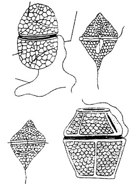
(
a)
(
b)
- Nucleus : Cells possess a relatively large and prominent nucleus Hypovalve known as mesokaryon. The interphase nucleus has condensed Longitudinal flagellum chromosomes which lack histones.
- Plastids : There are numerous discoid chloroplasts without pyrenoids. They are yellow-brown to dark-brown in colour due to presence of characteristic pigments – Chlorophyll a, c, α- carotene and xanthophylls (including dinoxanthin and peridinin).
- Reserve food : The reserve food material is starch or oil.
| (c) (d) | |
| (vi) Pusule : The cells possess an osmoregulatory organelle called pusule which superficially looks like contractile vacuole. | Fig : Some dinoflagellates (a) Glenodinium (b) Peridinium (c) Gynnodium |
(d) Gonyaulax
The cells posses mitochondria, ribosomes and Golgi bodies. They also possess mucilage bodies or vesicles below the cell membrane.
(3) Nutrition : In dinoflagellates it is mainly holophytic or photosynthetic. However, some forms are saprobic, parasitic, symbiotic or holozoic. For example, an colourless Blastodinium is parasite on animals.
(4) Reproduction
- Asexual reproduction : Dinoflagellates reproduce asexually through cell division or by the formation of zoospores and cysts.
The cell division starts from posterior end. During cell division, centromeres and spindle are not seen. The spindle is replaced by cytoplasmic microtubules. During mitosis, the chromosomes break up into pairs of romatids. The nuclear envelops and nucleolus persists during divison.
- Sexual reproduction : If it is occurs, is isogamous or anisogamous. Two cells conjugate by a conjugation canal where the two amoeboid gametes fuse to form a diploid zygote. Life cycle involves zygotic meiosis (e.g., Ceratium, Gymnodinium etc.) or gametic meiosis (e.g. Noctiluca).
Diatoms (Division-Bacillariophyta)
(1) Habit and Habitat : Most of the diatoms occur as phytoplanktons both in fresh and marine waters. A few forms occur as benthos the bottom of water reservoirs. Some are terrestrial and grow on moist soil. Diatoms constitute a major part of phytoplankton of the oceans. It is estimated that a 60 ton blue whale may have approximately 2 tons of plankton (mostly diatoms) in its gut.
(2) Structure
- Shape : The cells of diamots are called frustules or shell. They are microscopic, unicellular, photosynthetic organisms of various colours and diverse forms. They may be circular, rectangular, triangular, elongated, spindleshaped, half-moon shaped, boat-shaped or filamentous.
- Symmetry : They exhibit mainly two types of symmetry-radial symmetry as in centrales (e.g. Cyclotella) and isobilateral symmetry as in Pennales (e.g. Pinnularia).
- Cell wall : The cells of diatoms are called frustules. The cell wall is chiefly composed of cellulose impregnated with glass-like silica. It shows sculpturings and ornamentations. It is composed of two overlapping halves (or theca) that fit togather like two parts of a soap box. The upper half (lid) is called epitheca and the lower half (case) is called hypotheca.
- Flagella : Diatoms do not possess flagella except in the reproductive stage. They show gliding type of movement with the help of mucilage secretion. They float freely on the water surface due to presence of light weight lipids.
- Nucleus : Each cell has a large central vacuole in which a prominent nucleus is suspended by means of cytoplasimc strands. The cells are diploid (2 N). In case of centrales, the nucleus lies in the peripheral region.
- Plastids : The cells possess plate-like or discoid chromatophores (or chloroplasts). They contain Chlorophyll a, Chlorophyll c, carotenes, diatoxanthin, diadinoxanthin and fucoxanthin (chlorophyll b is absent).
- Reserve food : The reserve food material is oil and a polysaccharide – chrysolamisarin (or leucosin).
(3) Reproduction
- Asexual reproduction : The most common method of multiplication is binary fission (cell division) that occurs at night. In this process, each daughter individual retains one half of the parent cell and the other half is synthesized i.e. epitheca is retained and the hypotheca is synthesized. As a result, one of the two daughter individual is slightly smaller than the parent cell and there is gradual reduction in the frustule over the generations. However, the normal size is maintained by – formation of rejuvenescent cells (auxospores), growth of protoplast secreted from the frustule and secretion of new frustule of larger size.
- Sexual reproduction : Reproduction takes place by the fusion of gametes. Meiosis is gametic i.e. takes place during the formation of gametes. The diploid nucleus of parent cell divides by meiosis into 2 or 4 daughter cells. Out of 4 haploid daughter nuclei only one or two survive and rest degenerate. Thus they produce only one or two gametes. The gametes may come out by an amoeboid movement and fuse externally in pairs within a mucilaginous sheath to produce zygote. The diploid zygote then a transformed into auxospore.
Important Tips
- The term prokaryotes and eukaryotes were coined by Fott.
- In five kingdom system of classification, the main basis of classification is structure of nucleus.
- Gold Fuss give the term ‘Protozoa’.
- Ernst Haeckel proposed the term ‘Protista’.
- Photosynthetic protists fix about 80% of CO2 in the biosphere.
- Protistology : Study of protists.
- Monerology : Study of monerans.
- Slime moulds possess animal like as well as fungi like character.
- Euglenoids possess plant like as well as animal like characters.
- De Bary (1887) classified slime moulds as a animal and called them ‘Mycetozoa’.
- Macbrid coind the term ‘Myxomycetes’ (Slime moulds).
- Dinoflagellates, due to spinning caused by activity of transverse flagellum (in cingulum/annulus) and longitudinal flagellum (in sulcus), represent whorling whips.
- Dinoflagellates with bioluminescence/phosphorescence due to light producing protein luciferin are called fire algae. e.g. Noctiluca, Pyrocystis, Pyrodinium etc.
- Leeuwenhock (1674, 1675, 1681) was first to observe and sketch protozoan protists including Vorticella and Giardia.
- Acellular organisms do not contain cellular structure e.g., viruses or not considered as cells but as complete organisms e.g., protists.
- Wall-less multicellular protoplasm of acellular slime moulds having branched veins and with process of cyclosis are called phaneroplasmodium.
- Dinoflagellates symbionts in other protists and invertebrates are called zooxanthellae.
- Some dinoflagellates produce blooms or red tides. e.g. Gonyaulax, Gymnodinium etc.
- Silicon is present in the frustule of diatoms.
- Auxospones are formed by diatoms.
- Noctiluca dinoflagellate is called ‘night light’.
- Diatoms are emploid as a source of water glass or sodium silicate.
- Ganobacteria term was coined by IBCN (1978).
| Kingdom fungi. |
-
- Introduction : The science dealing with the study of fungi is called as mycology. The knowledge of fungi to mankind dates back to prehistoric times. Clausius, 1601 may be regarded as one of the earliest writers to describe fungi. Bauhin (1623) also included the account of known fungal forms in his book Pinax Theatric Botanica. The fast systematic account of fungi came from Pier Antonio Micheli (1729) who wrote ‘Nova Plantarum Genera’. He is described by some workers as founder or mycology. Linnaeus (1753) also included fungi included fungi in his ‘Species Plantarum’. Elias Fries (1821-31) gave a more detailed account of fungi in his ‘Silloge Fungorum’ in 25 volumes describing some 80,000 species of fungi. This work remains unparalleld even today.
- Thallus organization : The plant body of true fungi (Eumycota), the plant body is a thallus. It may be non-mycelial or mycelial. The non-mycelial forms are unicellular, however, they may form a pseudomycelium by budding. In mycelial forms, the plant body is made up of thread like structures called hyphae (sing. hypha). The mycelium may be aseptate (non-septate) or septate. When non-septate and multinucleate, the mycelium is described as coenocytic. In lower fungi the mycelium is non-septate e.g., Phycomycetae. In higher forms it is septate e.g., Ascomycotina, Basidiomycotina and Deuteromycotina. In some forms the plant body is unicelled at one stage and mycelial at the other. Their organization is sometimes described as dimorphic.
Holocarpic and Eucarpic : When the entire mycelium is converted into reproductive structure, the thallus is described as holocarpic. However, if only a part of it becomes reproductive, the thallus is called as eucarpic. The eucarpic forms may be monocentric (having a single sporangium) or polycentric (having many sporangia).
-
- Specialised formation : In higher forms the mycelium gets organised into loosely or compactly woven structure which looks like a tissue called plectenchyma. It is of two types :
- Prosenchyma : It comprises loosely woven hyphae lying almost parallel to each other.
- Pseudoparenchyma : If the hyphae are closely interwoven, looking like parenchyma in a cross-section, it is called as pseudoparenchyma.
In addition to above, the fungal mycelium may form some specialized structures as under :
-
- Rhizomorphs : Its a ‘root-like’ or ‘string-like’ elongated structure of closely packed and interwoven hyphae. The rhizomorphs may have a compact growing point.
- Sclerotia : Here the hyphae gets interwoven forming pseudoparenchyma with external hyphae becoming thickened to save the inner ones from desiccation. They persist for several years.
- Stroma : It is thick mattress of compact hyphae associated with the fruiting bodies.
- Cell organization : The cell wall of fungi is mainly made up of chitin and cellulose. While chitin is a polymer of N-acetyl glucosamine, the celulose is polymer of d-glucose. Precisely, the cell wall may be made up of cellulose-glucan (Oomycetes), chitin chitosan (Zygomycetes) mannan-glucan (Ascomycotina), chitin-mannan (Basidiomycotina) or chitin-glucan (some Ascomycotina, Basidiomycotina and Deuteromycotina). Besides, the cell wall may be made up of cellulose-glycogen, cellulose-chitin or polygalactosamine-galactan.
In higher fungi, where the mycelium is septate, the septa are of several types :
-
- Solid septum : It has no perforations.
- Perforated septum : It has several perforations.
- Acomycetean septum : It has a single large pore in the centre of the septum.
- Bordered pit type septum : It has a perforation in the septum resembling the bordered pit of tracheary elements.
(v) Dolipore septum : It has a single barrel shaped Plasma Endoplasmic
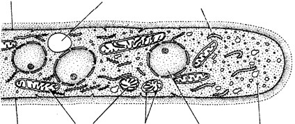 membrane Vacuole reticulum pore in the septum due to thickened rim. The pore has a cap of ER called parenthosome.
The cell wall is closely associated with the inner layer, the plasma membrane. In fungi, specialized structure called lomasomes are also found associated to the plasma
membrane. They appear to be as infoldings or invagination of Mitochondria Ribosome Nucleus Microbodies
Hyphal wall the membrane. Almost similar structures called
plasmalemmasomes are also found associated to the Fig : Diagrammatic electronmicrograph of a fungal cell membrane. The cell contains one or more well defined
eukaryotic nuclei. In fungi the nuclei show intranuclear mitosis which is sometimes referred to as karyochorisis. They also contains mitochondria, E.R., ribosomes, microbodies, lysosomes, vacuoles and crystals of reserve food particles (glycogen, lipid etc.). The cells lack golgi and chloroplast and therefore, chlorophyll and starch grains are also absent. However, a reddish pigment, neocercosporin has been isolated from the fungus Cercospora kikuchii. The vacuoles are bound by tonoplast. The genetic material is DNA.
(5) Nutrition : The fungi are achlorophyllous organisms and hence they can not prepare their food. They live as heterotrophs i.e., as parasites and saprophytes. Some forms live symbiotically with other green forms.
| ectoparasites occur on the surface of the host tissue whereas the endoparasites are found within the host tissue. The forms belonging to the third category are partly ecto- and partly endoparasites. In parasitic forms. The mycelium may | (c)
Fig : Parasitic hyphae and haustorial appendages : (a) Interacellular hyphae (b) and(c) Intercellular hyphae with |
- Parasites : They obtain their food from a living host. A parasite may be obligate or facultative. The obligate parasites thrive on a living host throughout their life. The facultative parasites are infact saprophytes which have secondarily become parasitic. While the above
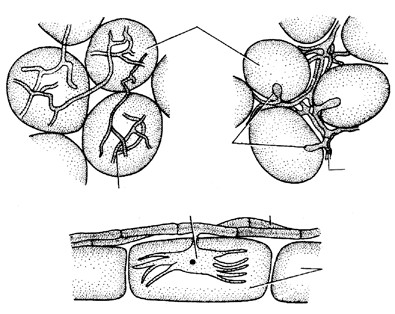
Host cells
Haustoria
Intracellular hyphae
Haustorium
Ectoparasitic hypha
Epidermal
cells
(
a)
(
b)
Intercellular hyphae classification is based on the mode of nutrition, however, on the basis of their place of occurrence on the host, the parasites can be classified as ectoparasite, endoparasite and hemiendoparasite (or hemiectoparasite). The
haustoria occur within the host cells (intracellular) or in between the host cells (intercellular).
Some forms produce rhizoids for absorbing food. The parasitic fungi produce appressoria for adhering to the host. For absorbing food, the obligate parasites produce haustoria. As a result, the plasma membrane of the host cell becomes convoluted but it does’nt break. The fungal cell wall also remains intact. The haustoria may be finger-like, knob-like or branched. Each haustorium is distinguishable into a base, stem and body.
- Saprophytes : They derive their food from dead and decaying organic matter. The saprophytes may be obligate or facultative. An obligate saprophyte remains saprophytic throughout it’s life. On the other hand, a facultative saprophyte is infact a parasite which has secondarily become saprophytic.
- Symbionts : Some fungal forms grow in symbiotic association with the green or blue-green algae and constitute the lichen. Here the algal component is photosynthetic and the fungal is reproductive. A few fungal forms grow in association with the roots of higher plants. This association is called as mycorrhiza. They are two types – Ectotrophic mycorrhiza and Endotrophic mycorrhiza e.g., (VAM).
(6) Reproduction : The fungi may reproduce vegetatively, asexually as well as sexually :
(i) Vegetative reproduction
- Fragmentation : Some forms belonging to Ascomycotina and Basidiomycotina multiply by breakage of the mycelium.
- Budding : Some unicelled forms multiply by budding. A bud arises as a papilla on the parent cell and then after its enlargement separates into a completely independent entity.
- Fission : A few unicelled forms like yeasts and slime molds multiply by this process.
- Oidia : In some mycelial forms the thallus breaks into its component cells. Each cell then rounds up into a structure called oidium (pl. oidia). They may germinate immediately to form the new mycelium.
- Chlamydospores : Some fungi produce chlamydospores which are thick walled cells. They are intercalary in position. They are capable of forming a new plant on approach of favourable conditions.
(ii) Asexual reproduction
- Sporangiospores : These are thin-walled, non-motile spores formed in a sporangium. They may be unior multinucleate. On account of their structure, they are also called as aplanospores.
- Zoospores : They are thin-walled, motile spores formed in a zoosporangium. The zoospores are of several types :
- Uniflagellate with whiplash type flagellum e.g., Allomyces.
- Uniflagellate with tinsel type flagellum e.g., Rhizidiomyces.
- Biflagellate with a tinsel type and a whiplash type flagella e.g., Saprolegnia.
- Biflagellate with two whiplash type flagella e.g., Plasmodiophora.
(c) Conidia : In some fungi the spores are not formed inside a sporangium. They are born freely on the tips of special branches called conidiophores. The spores thus formed are called as conidia. On the basis of development, two types of conidia are recognised namely thallospores and blastospores or true conidia.
Thallospores : In some forms the thallus itself forms spore like bodies called thallospores. The thallospore are of two types namely arthrospores and chlamydospores.
- Arthrospores : They are thinwalled spores formed in basipetal order e.g., Endomyces.
- Chlamydospores : Some of the hyphal cells are converted into thick walled chlamydospores. They may be terminal or intercalary e.g., Ustilago, Saprolegnia.
Blastospores : They develop on conidiophores in acropetal or basipetal succession. They are of two types –
- Porospores : When the blastospores develop by the balooning of the inner wall of conidiophore, it is called as porospore e.g., Alternaria.
- Phialospore : On the other hand, when the first conidium carries the broken parent wall of conidiophore and subsequent conidia possess a new wall, such basipetally formed conidia are called as phialospore e.g., Aspergillus.
Bi-celled conidia are formed in Trichothecium. In Fusarium it is possible to differentiate smaller microconidia from larger macroconidia. Sometimes the conidiophores form specialised structures as under :
Synnema or Coremium : Here the conidiophores get arranged in closely placed parallel plates.
Acervulus : It is a cushion-shaped mass of hyphae having closely packed conidiophores.
Sporodochium : It is also a cushion-shaped acervulus like structure having loosely arranged conidiophores.
Pycnidium : It is pitcher-shaped, embedded body which opens to exterior by a pore called ostiole. It is lined by conidiogenous hyphae. The conidia developing in pycnidia are often described as pycniospores.
(iii) Sexual reproduction : With the exception of Deuteromycotina (Fungi imperfecti), the sexual reproduction is found in all groups of fungi. During sexual reproduction the compatible nuclei show a specific behaviour which is responsible for the onset of three distinct mycelial phases. The three phases of nuclear behaviour are as under :
Plasmogamy : Fusion of two protoplasts.
Karyogamy : Fusion of two nuclei.
Meiosis : The reduction division.
These three events are responsible for the arrival of the following three mycelial phases :
Haplophase : As a result of meiosis the haploid (n) or haplophase mycelium is formed.
Dikaryotic phase : The plasmogamy results in the formation of dikaryotic mycelium (n + n).
Diplophase : As a result of karyogamy the diplophase mycelium (2n) is formed.
In some fungi plasmogamy, karyogamy and meiosis do occur in a regular sequence but not at specified time or points in life cycle. Such a cycle is described as parasexual cycle and phenomena celled parasexuality recorded by Pontecorvo and Roper.
The fungi reproduce sexually by the following methods :
- Isogamy : It involves fusion of two morphologically similar flagellate gametes.
- Anisogamy : Here the two gametes are motile but morphologically dissimilar. The larger gamete may be called as female and the smaller one as male.
- Heterogamy : It involves fusion of a non-motile female gamete (egg) with the motile male gamete (antherozoid). While the male gamete is formed inside the antheridium, the female is produced inside the oogonium. Both the sex organs are unicelled structure.
- Gametangial contact : It involves fusion of two gametangia. In lower forms the female gametangium is called as oogonium. The male gametangium is termed as antheridium. A contact develops in between the two gametangia and then the male nucleus is transferred into the female directly or through a tube.
- Gametangial copulation : In this case the fusion occurs in between the two gametangia. When it occurs in some holocarpic forms where the entire thallus acts as gametangium, the phenomenon is called as hologamy. In others, dissolution of cell wall in between the two gametagial brings about gametangial copulation.
- Spermatization : Here the uninucleate male gametes called spermatia are formed in special structures called spermogonia or pycnidia. The female gametangium is called as ascogonium which has a long neck called trichogyne. The spermatium attaches itself with the trichogyne and transfers the male nucleus, thus bringing about dikaryotisation.
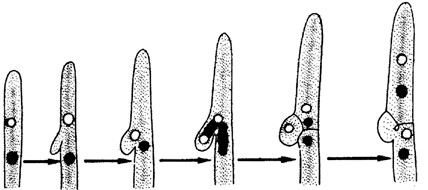 Somatogamy : In higher fungi there is reduction of sexuality to the maximum level. Here two hyphae of opposite strains are involved in fusion thus bringing about dikaryotization.
- Clamp connection : In Basidiomycotina, the dikaryotic cells divide by clamp connections. They were first observed by Hoffman, (1856) who
Dikaryotic Pouch Conjugate Septation Dissolution of cell formation division lateral septum
Fig : Various stages in the formation of clamp connections
named it as ‘Schnallenzellen’ (buckle-joints). A lateral pouch like outgrowth arises which projects downward like a hook. This pouch or clamp becomes almost parallel to the parent cell. The two nuclei now undergo conjugate division in such a way that one spindle lies parallel to the long axis of the cell and the other somewhat obliquely. As a result, one daughter nucleus enters into the clamp. Now, septae appear separating the clamp and the lower hyphal cell. The upper cell has both the nuclei. The clamp with a nucleus now fuses with the lower cell. The septum between the pouch and the lower cell is dissolved and thus the lower cell now contains both the nuclei of opposite strains. The entire process takes some 23-45 minutes.
- Heterothallism : Blakeslee, (1904) while working with Mucor sp. observed that in some species sexual union was possible between two hyphae of the same mycelium, in others it occured between two hyphae derived from ‘different’ spores. He called the former phenomenon as homothallism and the latter as heterothallism. Thus, the homothallic species are self-fertile whereas the heterothallic are self sterile. In heterothallic species the two ‘thalli’ are sexually incompatible. They are said to belong to opposite strains. Blakeslee designated them as + and – i.e., belonging to opposite strains or mating types. Whitehouse, (1949) differentiated the phenomenon into two categories as under :
- Morphological heterothallism : When male and female sex organs are located on different ‘thalli’ the heterothallism is said to be morphological. Such species are generally described as dioecious.
- Physiological heterothallism : In bisexual forms the two sex organs may be located on the same ‘thallus’ or on different ‘thalli’. In some forms the two sexes even when present on the same ‘thallus’ are unable to mate, the heterothallism is said to be physiological. Such forms are self-sterile as they need genetically different nuclei. Such nuclei are absent when the same ‘thallus’ forms the two sex organs. Heterothallic fungi may be bipolar or tetrapolar.
(9) Classification of fungi : It is based largely on the characteristics of the life cycle involved like.
Nature of somatic phase, kinds of asexual spores, kinds of sporangia, nature of the life cycle and presence or absence of perfect or sexual stage.
| Kingdom Fungi | |
Sub-kingdom
Gymnomycota Oomycota Eumycota
(Myxomycota) Mycelium Mycelium Slime Moulds now aseptate septate
Phycomycetes
excluded from fungi and
placed under protista
Zygomycetes
(
Conjugation fungi)
e.g. Rhizopus, Mucor
Mycophycophyta
(
Dual organisms)
Lichens,
e.g.,
Usnea, Parmelia
Deuteromycota
(
Fungi imperfecti)
Sexual reproducti
on absent,
e.g., Alternaria, Cercospora,
(Oomycetes
Algal fungi)
Phytophthora,
Albugo, Pythium
| Microsporum, Trichophyton. | Ascomycota
(Sac fungi) Aspergillus, Penicillium, Neurospora |
Basidiomycota
(Club fungi) Puccinina, Ustilago, Agaricus |
(10) Salient features of classes
- Phycomycetes (Oomycetes/Egg fungi) : It is also called lower fungi, mycelium is coenocytic. Hyphal wall may contain chitin or cellulose (e.g., Phytophthora). Asexual reproduction occurs with the help of conidiosporangia. Under wet conditions they produce zoospores. Under dry conditions, the sporangia directly function as conidia. Zoospores have heterokont flagellation (one smooth, other tinsel). Sexual reproduction is oogamous. It occurs by gametangial contact where male nucleus enters the oogonium through a conjugation tube. The fertilized oogonium forms oospore. e.g., Sapolegnia, Albugo (Cystopus), Phytophthora, Phythium, Sclerospora.
- Zygomycetes (Conjugation fungi) : Mycelium is coenocytic. Hyphal wall contains chitin or fungal cellulose. Motile stage is absent. Spores (Sporangiospores/aplanospores) are born inside sporangia. Sexual reproduction involve fusion of coenogametes through conjugation (Gametangial copulation). It produces a resting diploid Zygospore. On germination, each zygospore forms a germ sporangium at the tip of a hypha called promycelium e.g., Mucor, Rhizopus.
- Ascomycetes (Ascus : sac, mycete : fungus) : These are unicellular as well as multicellular fungi. In the latter, mycelium is septate. The asexual spores formed in chains are called conidia. The spores are formed exogenously, i.e. outside sporangium. They detach from the parent and form new mycelia. Sexual reproduction is through ascospores, which are formed endogenously (within the mycelium) in a sac like structure called ascus (pl. asci). The gametes involved in sexual reproduction are nonmotile compatible and are generally represented as + and –. The fusion of gametes is followed by reductional division that produces haploid ascospores. The fruiting body called ascocarp.
The ascocarp are of four types :
- Cleistothecium : It is an ovoid or spherical fruiting body which remains completely closed e.g., Aspergillus.
- Perithecium : It is a flask shaped fruiting body which opens by a single pore called ostiole. It is lined by sterile hyphae called paraphyses. The asci are also mixed with paraphysis e.g., Cleviceps.
- Apothecium : It is a saucer-shaped fruiting body. The asci constitute the fertile zone called hymenium e.g., Peziza.
- Ascostroma : It is not a distinct fruiting body. It lacks its own well defined wall. The asci arise directly with a cavity (locule) of stroma. It is also called as pseudothecium e.g., Mycosphaerella.
- Basidiomycetes : They are the most advanced fungi and best decomposers of wood. These are called club fungi because of a club shaped end of mycelium known as basidium. They have septate multinucleated mycelium. Septa possess central dolipores and Lateral clamp connections. The sexual spores called basidiospores are generally four in number. They are produced outside the body (exogenuous) unlike ascomycetes where they are endogenous. Two compatible nuclei fuse to form zygote, which undergoes meiosis and forms four basidiospores. The fruiting body containing basidia is a multicelular structure called basidiocarp. The common members are edible mushrooms (Agaricus). Smut and Rust.
- Deuteromycetes (Fungi inperfecti) : The group include all those fungi in which sexual or perfect stage is not known. Mycelium is made of septate hyphae. Asexual reproduction commonly occur by means of conidia.
(11) Economic importance
(i) Harmful aspects
(a) Crop diseases : Several important crop plants are destroyed due to fungi diseases. Some important ones are listed here under :
Some plant disease caused by fungi
| Disease | Crop | Causal organism |
| White rust of crucifers | Family Cruciferae | Albugo candida or Cystopus |
| Early blight of potato | Potato | Alternaria solani |
| Tikka disease of groundnut | Groundnut | Cercospora personata or C. arachidicola |
| Ergot disease of rye | Rye | Claviceps purpurea |
| Red rot of sugarcane | Sugarcane | Colletotrichum falcatum |
| Powdery mildew | Wheat | Erysiphe polygoni |
| Powdery mildew | Peas | Erysiphe graminis |
| Wilt of gram | Gram | Fusarium orthaceras |
| Bankanese disease and food rot of rice | Rice | Gibberella fujikuri |
| Leaf spot of oats | Oats | Helminthosporium avenae |
| Brown leaf spot of rice | Rice | Helminthosporium oryzae |
| Flag smut of wheat | Wheat | Urocystis tritici |
| Flag smut of oat | Oats | Ustilago avenae |
| Covered smut of barley | Barley | Ustilago hordei |
| Covered smut of oat | Oats | Ustilago kolleri |
| Smut of sugarcane | Sugarcane | Ustilago scitaminea |
| Loose smut of wheat | Wheat | Ustilago tritici |
| Late blight of potato | Potato | Phytophthora infestans |
| Club rot of crucifers | Cabbage | Plasmodiophora brassicae |
| Downy mildew of grapes | Grapes | Plasmopara viticola |
| Black rust of wheat | Wheat | Puccinia graminis-tritici |
| Brown rust of wheat | Wheat | Puccinia recondita |
| Yellow rust of wheat | Whet | Puccinia striiformis |
| Damping off of seedlings | Various seedlings | Pythium sp. |
| Blast of rice | Rice | Pyricularia oryzae |
| Grain smut of jowar | Jowar | Sphacelotheca sorghi |
| Wart disease of potato | Potato | Synchytrium endobioticum |
| Leaf curl of peach | Peach | Taphrina deformans |
- Diseases in human beings : Several diseases in human beings are found to be caused by fungi infecting different parts of the body. Some of them are given hereunder as :
| Disease | Causal organism | Place of infection |
| Athletes foot | Epidermophyton floccosum | Foot |
| Ring worm | Trichophyton sp., Microsporum sp., Epidermophyton sp., Myxotrichum sp. | Skin |
| Moniliasis | Candida albicans | Nails |
| Aspergillosis | Aspergillus niger, A. flavus, A. terrus | Lungs |
| Torulosis | Cryptococcus neofomans | Lungs, CNS |
- Spoilage of food : Some forms like Rhizopus, Mucor, Aspergillus, Cladosporium grow on food articles and spoil them. Cladosporium grows even at a temperature of – 6°C.
- Mycotoxins : Some fungi produce toxic metabolites which cause diseases in human beings. They usually contaiminate cereals and oil seed crops. Four types of mycotoxins are generally identified :
- Aflotoxins : e.g., Aflotoxin B1, B2, M1, M2, G1, G2; They are produced mainly by Aspergillus flavus and A.
parasiticus. They are well know for their carcinogenic effect.
- Zearalenone : It is produced by Fusarium sp.
- Ochratoxins : They are produced by Aspergillus and Pennicillium sp.
- Trichothecenes : They are produced by fungi like Cephalosporium, Fusarium etc.
- Poisonous fungi : Some fungi are extremely poisonous e.g., Amanita phalloides (‘death cup’). A. verna, Boletus satanus. Forms like Coprinus, Psilocybe are less poisonous. The fungus Amanita phalloides produces toxins like α-amanitin, phalloidin etc. which are very poisonous.
- Ergotism :The fungus causing ‘ergot’ disease of rye (Secale) is Cleviceps purpurea. It contains many poisonous alkaloids in their sclerotia. It causes poisoning in human beings. It’s acute condition is called as ‘St. Anthony’s fire‘.
- Hallucinogenic drugs : The hallucinogenic drug LSD (Lysergic acid Diethylamide) is extracted from Cleviceps purpurea as also from Inocybe. Besides, the mushroom Amanita muscaria is also hellucinogenic.
- Rotting of wood : Rotting of wood is caused due to degradation of lignin and cellulose. It is brought about fungi like Polyporus sp., Fomes sp. and Ganoderma sp., Forms like Fusarium, Penicillium leave stains on the wood.
- Allergies : Spores of Mucor, Aspergillus, Penicillium, Puccinia etc., present in the atmosphere cause allergies.
- Deterioration of articles : Forms like Aspergillus, Cladosporium, Rhizopus, Chaetomium, Alternaria deteriorate cork, rubber, leather, textile and even plastics.
(ii) Useful aspects
- Food : Forms like Agaricus bisporus, Morchella esculenta, Lentinus edodes, Clavatia gigantia, Volvariella volvacea are edible. The yeast Saccharomyces cerevisiae is used for making ‘yeast cake‘. When mixed with cereal flour, the yeasts produce a preparation called incaparina. The Single Cell Protein (SCP) obtained from yeasts, Penicillium, Fusarium etc. are used as substitute of protein food, Rhizopus oligosporus, when processed with soybeans yield a food preparation called ‘tempeh’. It has high protein contents.
- Flavoring of food : Penicillium roquefortii and P. camemberti are employed for flavoring cheese.
- Brewing and baking : Yeasts are generally used in bakeries and breweries. The sugars are fermented by yeasts into alcohol and CO2. While the former in main product of breweries CO2 is mainly useful in bakeries.
- Organic acids : Several organic acids are commercially produced by fungi, some of which are given hereunder :
| Organic acids | Source |
| Citric acid | Aspergillus niger |
| Gallic acid | Penicillium glaucum |
| Gluconic acid | Aspergillus niger, Penicillum purpurogenum |
| Fumaric acid | Rhizopus stolonifer, Mucor sp. |
| Lactic acid | Rhizopus nodosus |
| Kojic acid | Aspergillus flavus |
| Oxalic acid | Aspergillus niger |
| Acetic acid | Candida sp. |
- Antibiotics : The antibiotics are chemicals produced by living organisms that kill other living organisms. The first known antibiotic is penicillin that was extracted from Penicillium notatum by A. Fleming, (1944). Raper (1952) also extracted the same antibiotic from P. chrysogenum. Besides, several other antibiotics have been extracted since then.
| Antibiotics | Source |
| Griseoflavin | Penicillium griseofulvum |
| Citrinin | P. citrinum |
| Cephalosporin | Acremonium sp. |
| Ramycin | Mucor ramannianus |
| Proliferin | A. proliferans |
| Jawaharin | A. niger |
| Patulin | Aspergillus clavatus |
| Fumigatin | Aspergillus fumigatus |
| Viridin | Gliocladium virens |
| Penicillin | Penicillium chrysogenum, Penicillium notatum |
| Trichothecin | Trichothecium roseum |
| Campestrin | Agaricus campestris |
| Frequentin | Aspergillus cyclopium |
| Chaetomin | Chaetomium cochloids |
| Ephedrin | Yeasts |
- Other chemicals : Various chemicals have been obtained from different kinds of fungi. Yeast are good source of glycerol and enzymes like zymase, invertase and lipase. Cellulases are obtained from Aspergillus. Some alkaloids are also obtained from fungi e.g., Ergotinine, Ergotetrine and Ergobasine from Cleviceps purpurea. Gibberellins (plant hormones) are obtained from Gibberella fujikuroi. Another hormone, trisporic acid is obtained from Mucor mucedo.
- Biological assays : The fungi can detect the presence of certain chemicals present in the medium even in traces e.g., Aspergillus niger for Mn, Pb, Zn, Cu, Mo etc.
- Vitamins : Various vitamins have been obtained from different kind of fungi.
| Vitamins | Source |
| Vitamin A | Rhodotorula gracilis |
| Vitamin B12 | Eremothcium ashbyii |
| Thiamine B1 | Saccharomyces cerevisiae |
| Riboflavin B12 | Saccharomyces cerevisiae |
(i) Other uses
- Nitrogen fixation by yeasts like Saccharomyces and Rhodotorula.
- Production of latex by Mycena galopus.
- Soil building by Rhizopus, Cladosporium, Aspergillus etc.
- Along with bacteria, the fungi work as decomposers.
- Biological control of growth of hyperparasites like insects, nematodes, bacteria and even other fungi.
- Neurospora is a good research material for geneticists and Physarum for molecular biologists for the study of DNA.
Important Tips
- Fungus : The term was used by Gaspard Bauhin (1560–1624).
- Father of Mycology : Pier Antonio Micheli. In 1729 he wrote ‘Nova Genera Plantarum’ in which 900 fungi were described.
- Father of Systematic Mycology : E.M. Fries (1794–1878). He wrote ‘Systema Mycologicum’ in three volume.
- Father of Modern Mycology and Plant Pathology : H.A. de Bary.
- Father of Indian Mycology and Plant Pathology: E.J. Butler.
- Smallest Fungus : Yeast with a size of 3–15 µm × 2–10 µm.
- Largest Fungus : Lignocolous Shelf Fungus/Bracket Fungus Ganoderma applanatum (fruiting body 60 cm across). Glant
Puffball/Clavatia is 90–120 cm across. It possesses anticancer properties.
- Millardet discovered fungicide Bordeaux mixture. Which is solution of copper sulphate and calcium hydroxide (CaSO4 + Ca(OH)2).
- Pseudogamy : Fusion between unrelated cells.
- Pedogamy : Fusion between mature and immature cells.
- Adelphogamy : Fusion between mother and daughter cells or sister cells.
- Non-ciliated spores called ‘aplanospores’.
- Bipolar heterothallism found in Mucor and Rhizopous.
- Reserve food material of fungi is glycogen and oil bodies.
- A fungus which requires only one single host for complition of its life cycle is called ‘autoecious’.
- Phycomycetes are called algal fungi or lower fungi.
- Fungi inhabiting wood are known as epixylic.
- Aspergillus secretes toxin during storage conditions of crop plants. The hyphae of this fungus are septate and uninucleate.
- Ascomycetes are our worst fungus enemies.
- Neurospora (an ascomycete) is known as Drosophila of plant kingdom.
- Peziza and Helvella are coprophilous fungi (grow on dung).
- In higher Ascomycota the ascus develops indirectly from the tip of a bi-nucleate ascogenous hypha by becoming curved forming a crozier.
- Emperor Claudius Caesar was murdered by his wife by giving extract of toad stool fungus – Amanita phylloides which stops m-RNA synthesis, therefore it is called ‘Caesar’s Mushroom’.
- About 2300 antibiotics have been discovered so far from various fungi. Of these, some 123 have been extracted from Penicillium and 115 from Aspergillus.
- Destruction of potato crop by Phytophthora infestans in Ireland in 1845–49.
- Decrease in the yield of coffee in Srilanka from 42 m. kg. to 3 m. kg due to Hemileia vastatrix.
- Destruction of 5 million elm trees in England in 1967–77 by Ceratocystis ulmi.
- Destruction of 50% maize plants in USA (1970) due to infection of Helminthosporium maydis.
- Famous famine of Bengal in 1942–43 was due to destruction of rice crop by Helminthosporium oryzae.
- Few leading Indian mycologists are : C.V. Subramaniam, T.S. Sadasivan, K.C. Mehta and B.B. Mundkar.
- The fungi which grow on tree are called as lignicolous fungi e.g. Polyporus.
- Keratimorphic fungi appear on nails, feathers, hairs, hoofs etc.
- Hydnum from order agaricales is called tooth fungus.
- Balanced Parasites : Parasites which draw nourishment from hosts without killing or weakening them are called balanced parasites.
The parasites which bring about disease and destruction of the hosts are known as destructive parasites.
- Biotrophic Parasite (Gaumann, 1946). Absorbs nutrients from living host/cells.
- Nacrotrophic Parasite (Gaumann, 1946). Kills host cells for obtaining nourishment.
- Luminescent Fungi. They make wood/leaves/soil luminescent at night. Luminescent parts are pileus in Panus and Pleurorus species, fruiting body and mycelium in Clitocybe illudens.
- Acetyl Glucosamine/Chitin. (C22H54N4O21)n.
- Vegetative Vultures. Saprophytic fungi have been called vegetative vultures by Rolfe and Rolfe (1926) as they function as natural scavengers.
- Primary Host. Host in which the parasite becomes sexually mature. In stem rust the primary host is Wheat (karyogamy occurs) while alternate/secondary host is Barberry.
- Penicillin – Penicillium chrysogenum (initially from P. notatum) first antibiotic drug called wonder drug (discovered by Fleming, 1929).
- Species of Morchella are commonly known as ‘morels’, ‘sponge mushrooms’ or ‘gucchi’.
- The species of Polyporus are commonly called ‘bracket fungi’ or ‘self fungi’.
- Phytotoxin are secreted by plants in response to fungal reactions. They are generally phenolic compound.
- Coprophilou fungi grow on dung e.g., Pilobolus crystallinus.
- Fungi can be stained by cotton blue.
- Deuteromycetes is also known as ‘Fungal waste Basket’.
- The edible part of mushroom is basidiocarp.
| Rhizopus/Mucor. |
Systematic position
| Kingdom | – | Plantae |
| Sub kingdom | – | Thallophyta |
| Division | – | Mycota |
| Sub division | – | Eumycotina |
| Class | – | Zycomycetes |
| Order | – | Mucorales |
| Family | – | Mucoraceae |
| Genus | – | Rhizopus (For UP CPMT Students Only) |
Mucor (For MP PMT Students Only)
- Habitat : They are cosmopolitan and saprophytic fungus, living on dead organic matter. Rhizopus stolonifer occur very frequently on moist bread, hence commonly called black bread mold Mucor is called dung mold. Both are called black mold or pin mold because of black coloured pin shead like sporangia. Besides, it appears in the form of white cottony growth on moist fresh organic matter, jams, jellies, cheese, pickles, etc.
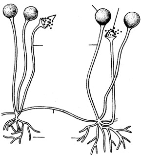
Sporangium
Sporangio-
spores
Sporangiophores
Stolon
Rhizoidal
hyphae
Columella
- Structure : The vegetative body or thallus consist of well branched, aseptate and multinucleate (coenocytic) mycelium on the surface of substratum. The mycelium is almost white when young but becomes blackish during reproductive phase. The mature mycelium is distinguishable into three types of hyphae :
- Stoloniferous hyphae : These hyphae grow horizontly on the surface of substratum. They are relatively stout and less branched than Fig : Rhizopus – Habit sketch showing stolon, rhizoidal hyphae and sporangiophores other hyphae. Certain portions of the stolons called nodes, give out rhizoids and sporangiophores.
- Rhizoidal hyphae : They arise in clusters from the lower side of each node and are repeatedly branched. The rhizoids penetrate the substratum and serve as anchors for the superficial mycelium. These hyphae secrete enzymes like amylase and maltase into the substratum and absorb the digested food.
- Sporangiophores : They are erect, aerial, unbranched reproductive hyphae that arise in clusters from the upper side of each node. Each sporangiophores develops single terminal sporangium which is filled with spores.
In Mucor there is no such distinction. In Mucor, the hyphae develop singly. There is no holdfast or apparant node. The hyphal wall is made up of chitin-chitosan and other polysaccharides. Inner to the chitin wall is a thin layer of plasma membrane. The granular protoplasm has many nuclei, glycogen and oil droplets, mitochondria, endoplasmic reticulum and ribosomes.
(3) Reproduction : They reproduces by vegetative, asexual and sexual methods.
- Vegetative reproduction : It takes place by fragmentation. If stolon breaks accidentally into small segments, each part grows into a new mycelium.
- Asexual reproduction : It occurs by three types of non-motile mitospores, sporangiospores, chlamydospores and oidia.
- Sporangiospores : The sporangiospores are also called aplanospores. They are thin walled, nonmotile, multinucleate spores formed in a sporangium. A vertically growing mycelium acts as sporangiophore. Its tip now shows accumulation of food and nuclei. The tip swells up into a vesicle which gradually enlarges. Soon the protoplasm gets demarkated into an outer dense region having many nuclei and inner vacuolated region having only a few nuclei. A septum now appears separating the outer sporangium from the inner columella. The protoplasm of the sporangium now shows formation of spores by cleavage which starts from the periphery. The sporangium dehisces irregularly due to collapse of columella and the spores are dispersed. The spores germinate under favourable conditions to form the new mycelium.
- Chlamydospores : These are the perennating spores formed when the fungus starts facing dry conditions. The protoplasm of hyphae collects at certain places, rounds off accumulates a lot of food materials and develops thick wall to become chlamydospores. They tide over the unfavourable conditions and germinate to produce new mycelia as soon as they get favourable conditions.
- Oidia : In liquid, sugary and acidic medium the hyphae form small rounded cells called oidia. They multiply by budding like yeast. The budded state is called torula stage. It takes part in alcoholic fermentation. On
transfer to a suitable solid medium, each oidium forms a new mycelium.
(iii) Sexual reproduction : Sexual reproduction takes place by conjugation between two multinucleate but single celled gametangia. The gametes are isogamous and non-motile.
The species of Rhizopus may be heterothallic (R.Stolonifer) or homothallic (R. sexualis). But mostly heterothallic in both Mucor and Rhizopus. In homothallic species sexual union in brought about between two hyphae of the same mycelium whereas in heterothallic species it occurs between two hyphae derives from different compatible strains i.e., positive (+) and negative (–).
In R. stolonifer the sexual reproduction occurs between two hyphae of opposite strains. It has been suggested by Burgeff (1924) and Mesland et al. (1974), that when two compatible strains approach each other, the following three reactions occur in members of Mucorales :
- Telemorphic reaction : The hyphae which form the progametangia are called zygophores. In this reaction club shaped zygophores are formed. The zygophore formation is induced by the hormones trisporic acids B and C.
- Zygotropic reaction : It involves the growth of zygophores from +ve and –ve strains towards each other. The growth of zygophores occurs as a result of some chemotropic response.
- Thigmotropic reaction : The changes taking place as a result of fusion or contact between the two zygophores, such as gametangial
(–)
(G)
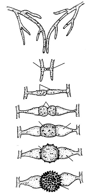
Zygophore
Gamet angia
Fusion cell
Suspensor
Developing
zygospore
Suspensor
Zygospore
(
F)
(
E)
(
D)
(
C)
(
B)
(
A)
Zygophore
Progametangia
(+)
Mycelium
Fig : (A)-(G) Stages in the sexual reproduction upto the formation of zygospore
fusion and septation, are controlled by this reaction.
The two mycelial branches growing towards each other are called progametangia. Their tips become rich in food and nuclei. They enlarge and come in contact each other. A septum is laid down separating the terminal gametangium from the proximal suspensor. The gametangium has dense cytoplasm and many nuclei whereas the suspensor has vacuolated cytoplasm with fewer nuclei. Each gametangium behaves as an aplanogamete or coenogamete. The two gametangia fuse with each other. Plasmogamy is followed by pairing of nuclei of opposite strains. The unpaired nuclei degenerate. This is followed by karyogamy. The zygospore so formed develops a dark coloured thick wall and undergoes rest. It is also believed that karyogamy is delayed till the germination of zygospore.
On the arrival of favourable conditions the zygospore germinate. The outer wall ruptures and the inner protrudes out in the form of promycelium. The promycelium grows vertically upward and forms a terminal germsporangium. It is generally believed that meiosis occurs in the germ sporangium. Each diploid nucleus forms four haploid nuclei, of which three degenerate. However, according to Cutter (1942) and Laane (1974), early nuclear divisions in the promycelium are meiotic. The germsporangium shows formation of thin walled spores which are initially uninucleate. Later on, by simple mitotic divisions they become multinucleate. The germsporangium ruptures irregularly releasing the spores which germinate to form a new mycelium. Occasionally, failure of gametangial copulation results in parthenogenous development of zygospores which are called
azygospore
(parthenospores)
.
Germination
Spore n
Melosis
Germsporangium
2
n
Promycelium
n
2
Zygospore
n
2
Rhizopus
& Mucor (n)
Progametangium (+)
Gametangium (+)
Plasmogamy n +
Karyogamy 2n
Zygophore (–) n
Progametangium (–)
Gametangium (–)
Zygophore (+)
Sporangiophore
Sporangium
Sporangiospores
(
Aplanospores)
Germination
Asexual
Chlamydospor
Sexual
Fig : Graphical representation of life cycle of rhizopus and mucor sp.
(4) Economic importance
- Spoilage of food : Exposed bread and other food particles are spoiled by Rhizopus and Mucor sp.
- Soft rot : Rhizopus species attack sweet potato, apple and strawberry producing soft rot or leak disease. Germinating maize grains are also attacked.
- Mucormycosis : Mucor pusillus and M. ramosissimus may attack internal human organs including lungs alimentary canal and nervous system
- Fermentad foods : They are prepared from rice and soyabean with the help of Rhizopus and Mucor e.g
Sufru,
- Chemicals : Citric acid prepared by Mucor from molasses, fuimaric acid and cortisone by Rhizopus stolonifer, Lactic acid by R. stolonifer and R.nodosus and alcohol by R. oryzae and M. javanicus.
- Antibiotic : Ramysin is produced by Mucor ramannianus.
- Waste water treatment : Growth of Mucor arrhizus removes heavy metal contamination of water.
| Yeast. |
Systematic position
| Division | – | Mycota |
| Sub division | – | Eumycotina |
| Class | – | Ascomycetes |
| Order | – | Endomycetales |
| Family | – | Saccharomycetaceae |
| Genus | – | Saccharomyces (Yeast) (For UP CPMT Students Only) |
- Habitat : Yeast is a saprophytic fungus found on substratum which is rich in sugars e.g. sugarcane, juice, fruits (palms, grapes), milk, etc. Some species are found on animal excreta.
- Structure : Yeast was first described by Antony Von Leeuwenhock in 1680. The yeast plant consists of a single cell which is very small and either spherical or oval in
Cell wall shape. However, under favourable conditions they grow rapidly
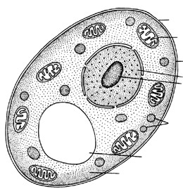
Vacuole
Cytoplasm
Mitochondrion and form false mycelium or pseudomycelium. Individual cells Cytoplasmic membrane are colourless but the colonies may appear white, red, brown, Nucleus creamy or yellow. The single cell are about 10µm in diameter. It Nucleolus is enclosed in a delicate membrane which is not made up of Food particles fungal cellulose but is a mixture of two polysaccharides known as mannan and glycogen. The cytoplasm in granular in appearance due to the
Fig : Electron micrograph of single yeast cell presence of droplets or granules of fat, glycogen and volutin. The volutin is nothing but nucleic acid. The glycogen is the chief reserve food material and its bulk increases during alcoholic fermentation and at times it may form as much as 30% of the weight of the yeast cell.
Yeasts are facultative aerobes i.e. they are anaerobes but can also survive under aerobic conditions and respire aerobically as well. The yeast cells secrete extracellular enzyme zymase which converts complex sugars into simple soluble sugars that can easily be assimilated.
(3) Reproduction : Yeast reproduces by vegetative or asexual and sexual methods.
(i) Vegetative reproduction : Yeast reproduce vegetatively either by fission or by budding
- Budding : It is the common method of reproduction in budding yeasts (e.g., Saccharomyces) under favourable conditions (i.e., when growing in sugar solution). During this process a small bud like out growth appears at one end of the parent cell which gradually enlarges in size (unequal division of cytoplasm takes place) The nucleus enlarges and divides amitotically into two daughter nuclei. One daughter nucleus goes into the bud and the other remains in the parent cell. The nuclear membrane persists throughout the nuclear division.
Daughter
buds
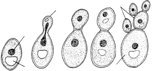
Mother cell
Vacuole
Developing
bud
Nucleus
(A) (B) (C) (D) (E)
Fig : Budding in yeast
The vacuole almost disappears at this stage. Gradually the bud becomes almost of the same size as the parent cell. Then a constriction appears at the base of bud and a separating wall (made up of chitin) is laid down. Sometimes a bud may produce another bud over it which is still attached to parent cell forming a false mycelium or pseudomycelium.
- Fission : It is a common method of reproduction in fission yeasts (e.g., Schizosaccharomyces). During fission the parent cell elongates and its nucleus divides into two daughter nuclei. The two nuclei separate apart. It is followed by a transverse cytokinesis by formation of a transverse septum which develops centripetally. The two cells separate apart and behave as uninucleate vegetative thalli.
Vacuole Dividing cell
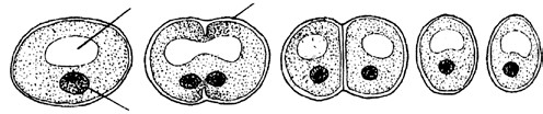
Divided cells
Nucleus
(A) (B) (C) (D)
Fig : Fission in yeast
(ii) Sexual reproduction : Sexual reproduction in yeasts takes place during unfavourable conditions, particularly when there is less amount of food.
The sex organs are not formed in yeasts and the sexual fusion occurs between the two haploid vegetative cells or two ascospores which behave as gametes. The two fusing gametes are haploid and may be isogamous or anisogamous. Such kind of sexual reproduction is called gametic copulation. It is the best example of hologamy i.e., the entire vegetative thallus is transformed into reproductive body. The sexual fusion leads to the formation of diploid zygote. The zygote behaves as an ascus and forms 4 – 8 haploid ascospores. These liberate and function as vegetative cells.
Guilliermond (1940) has recognised three types of life cycle in yeasts.
- Haplobiontic life cycle : This type of life cycle is common in Schizosaccharomyces octosporous, a homothallic species. It’s cells are haploid and they multiply by fission. Two haploid cells now act as gametangia and produce tiny protuberances towards one another. They fuse with each other to form a small conjugation canal or copulation tube. The nuclei of the two gametangia move into the tube and fuse to form a diploid zygotic nucleus. The zygote so formed behaves as ascus mother cell. It undergoes meiosis and then mitosis to form eight haploid nuclei which organise into eight ascospores. The ascus ruptures releasing the ascospores. They enlarge and behave as independent organisms.
- Diplobiontic life cycle : This type of life cycle is found in Saccharomyces ludwigii. The cells of this yeast are diploid and they multiply by budding. Ultimately a diploid cell functions as ascus, forms four ascospores which fuse in pairs. Each zygote grows into sprout mycelium from which vegetative cells develops as buds.
- Haplodiplobiontic life cycle : This type of cycle is found in Saccharomyces cerevisiae in which haploid and diploid both types of generations are found. The haploid nucleus of the ascus divides in two by mitosis and then followed by meiosis resulting in four small nuclei, the two of which being of (+) strain and the remaining two of the (-) strain. Thus four haploid ascospores are formed. The ascus wall ruptures releasing the four ascospores which start budding and produce new yeast cells. These cell are of (+) and (–) strains and function as gametes. When the two cells of different strains come together they fuse to form large yeast cell. Thus the alternation of generation takes places between the haploid – diploid generations.
Fission
n
Vegetative
cells n
Zygote 2n
Meiosis
Ascus
Ascospores
Vegetative
cells 2n
Vegetative cells
Ascospores
Sprout
cells
Zygote
Ascus n
Meiosis
Vegetative
cells n
Ascospores
n
2
cells
Ascus
Meiosis
Fig : Yeast graphical representation of life cycles
(
A) Haplobiontic, (B) Diplobiontic, (C) Haplodiplobiontic
Zygote
(
a)
b)
(
(
c)
In addition to above, in Schizosaccharomyces pombe, two adjoining sister cells fuse and this phenomenon is called adelphogamy. In some yeasts e.g. Debaryomyces, the mother and daughter cells fuse to form the zygote and this phenomenon is called pedogamy.
(4) Economic importance
(i) Useful activities
- Baking industry : Yeast are used in manufacture of bread. Kneaded flour is mixed with yeast and allowed to ferment. Yeast convert starch into sugars and sugar into CO2 and alcohol with help of enzyme zymase CO2 is released when effervescence takes place due to which bread become spongy and gets swollend and is of light weight.
- Brewing industry : Brewer’s/Beer yeast is Saccharomyces cerevisiae and wine yeast is Saccharomyces ellipsoidens. They perform alcoholic fermentation.
C H O6 12 6 →Yeast C H OH2 5 + 2CO2
Glucos e Zymase ethyl alcohol
(c) Food yeast : Yeast from brewing industry is harvested and used as food yeast. It is rich in protein and Bvitamins (Riboflavin) Special food yeasts are Torulopsis (protein), Endomyces (fat) and Cryptococus (both).
(ii) Harmful activities
- Fermentation of fruits and fruit juices by yeast cells makes their taste unpleasent.
- Parasitic species of yeast like Nematospora causes diseases in tomato, cotton and bean.
- Parasitic yeast cause diseases in human beings (e.g. cryptococcois, blastomycosis and torulosis).
| Albugo. |
Systematic position
| Kingdom – | Plantae |
| Sub kingdom – | Thallophyta |
| Division – | Mycota |
| Subdivision – | Eumycotina |
| Class – | Oomycetes |
| Order – | Peronosporales |
| Family – | Albuginaceae |
| Genus – | Albugo (For Rajasthan PMT Students Only) |
- Habitat : Albugo is an obligate parasite and grows in the intercellular spaces of host tissues. It is parasitic mainly on the members of families Cruciferae, Compositae, Amarantaceae and Convolvulaceae, The disease caused by this fungus is known as white rust or white blisters which is prevalent all over the world. The fungus forms shiny white patches on the leaves, mostly on their lower surfaces, stems and petioles. The fungus also causes hypertrophy and deformation in affected parts.
The most common and well known species is Albugo candida which attacks the members of the mustard family (Cruciferae). It is commonly found on Capsella bursa – pastoris (Shepherd’s purse) and occasionally on radish mustard, cabbage, cauliflower, etc. The reserve food is oil and glycogen.
- Structure : The plant body of the fungus is mycelial and eucarpic. The mycelium is intercellular, branched, aseptate and multinucleate (coenocytic). The mycelium produces finger like or globular haustoria which enter into the host cells to absorb the food material. The mycelial wall is made up of cellulose-glucan. The cells show characteristic fungal eukaryotic organization. The parietal layer of cytoplasm also contains oil globules.
Fungal cytoplasm
 Glycogen Fungal cell wall Lamasomes
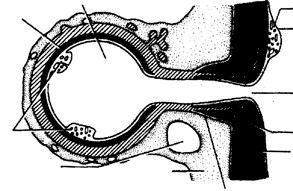
Host cell
vacuole
bodies Mycelium Fungal
plasmamembrane
Haustorium Host cell Fungal vacuole cytoplasm
Sheath
Host cell Lomasomes Fungal
Oil drops Host cell wall
-
- cytoplasm Host plasma
- membrane
Fig : Albugo – (A) Intercellular mycelium with knob like
haustoria, (B) Ultra structure of haustorium (diagrammatic)
- Reproduction : The fungus Albugo reproduces asexually as well as sexually.
(i) Asexual reproduction : Asexual reproduction take place by the formation of sporangia or conidia. The fungal mycelium collects below the host epidermis and forms sporangiophores (conidiophores). These are clubshaped and multinucleate. The sporangiophores cut sporangia in basipetal manner, that is, the oldest and first formed are at the top and the youngest and last formed are at the base. The sporangia remain attached in the form of chains .The walls between the sporangia fuse to form a gelatinous disc-like structure called disjunctor. Due to formation of numerous sporangia, the epidermis of the host is raised in the form of a postule.
Finally the disjunctors are dissolved in water and sporangia are set free. They appear as white powdery mass on the host tissue. These smooth, rounded structures are disseminated by wind. Germination of sporangia occurs when they reach a suitable host. The mode of germination depends on the availability of water or even moist air.
- If water is available sporangia forms About 4 – 12 (usually eight) zoospores within two minutes of their formation. The zoospores are kidney shaped and laterally biflagellate (unequal). The zoospores move into a vesicle before they are released. After a swimming period, the zoospores come to rest, encyst and germinate by germ tubes. The germ tubes enter into the host and produce an intercellular mycelium.
- If water is not available, the sporangia germinate directly by forming germ tubes.
(ii) Sexual reproduction : The sexual reproduction is oogamous type and takes place with the help of antheridia and oogonia. The antheridia and oogonia are multinucleate in the beginning but become uninucleate by disorganization of nucleri. the antheridia are club- shaped and the oogonia are globular. Both sex organs develop terminally on the hyphae. There is a single egg (oosphere) surrounded by periplasm. At the time of fertilization, a receptive papilla develops on one side of the oogonium through which the fertilization tube enters into the oogonium. Inside the oogonium the male nucleus fuses with the egg nucleus. The diploid zygote develops a warty wall and becomes the oospore. The diploid nucleus undergoes meiosis, followed by several mitotic divisions. After a period of rest, the oospore germinates and produces reniform, biflagellate zoospores. The zoopores are first released into a vesicle and then to the outside. They swim for some time, encyst and then germinate to form germ tubes. Most part of the life cycle of Albugo is gametophytic. The sporophytic phase is limited only to the oospore stage.
According to Sansome and Sansome (1974), the species A. candida is heterothallic and meiosis occurs inside the gametangia (antheridia and oogonia) and not in the oospore. Thus the vegetative mycelium of Albugo is diploid.
Germination
Zoospore
n
2
Oospore
2n
Karyogamy
Albugo (2n)
Meiosis
Sporangiophore
conidiophore) 2n
(
Sporangium
conidium) 2n
(
Zoospores 2n
germination
Asexua
Antheridum 2n
Fertilization
tube n
Male nucleus n
Oogonium 2n
Egg n
Female nucleus n
Sexual
Encystation
Fig : Graphical representation ofl ife cycle of rhizopus and mucor sp.
| Lichen. |
A lichen is structurally organised entity consisting of the permanent association of a fungus and an alga. The fungal component of a lichen is called mycobiont (mostly ascomycetes) and the algal component is called phycobiont (mostly blue green alga). Both mycobiont and phycobiont are associated in symbiotic union in which the fungus is more predominant and alga is subordinate partner. The fungus provides the structural covering that protects alga from unfavourable conditions, i.e., drought, heat etc. It also traps moisture from the atmosphere and anchors the lichens to a rock, tree bark, leaves and other similar supports. The alga prepares organic food (e.g., mannitol) by the process of photosynthesis from carbon dioxide. If the algal component is a cyanobacterium (blue green alga), it fixes atmospheric nitrogen in addition to preparation of food. The relationship between the two is that of consortium, symbiosis or mutualism. Ahmadjian (1963) considers fungus to be a controlled parasite. The phenomenon is called helotism.
- Habitat : Lichens are cosmopolitan in distribution.Their growth is very slow. Some lichens growing in arctic regions are believed to be 4500 year old. The lichens which grow on stones are called saxcoles (e.g., Dermatocarpon) and those growing on barks are called corticoles (e.g., Usnea). A few liches are aquatic (e.g., Peltigera, Verrucaria margacea). The lichens generally do not grow near smoky industrial areas where atmosphere is polluted. Cladonia rangiferina, commonly known as reindeer-moss grows luxuriantly in tundra region and form the food of animals like the reindeer and caribou (musk ox). Some of the common Indian genera are: Cladonia, Parmelia, Usnea, Physcia, Anaptychia, Lecidia, etc. Lichens are highly pigmented. They may be bluish, green, grey, yellow, orange, red and brown in colour. Some are white (Gyrophora).
- Classification : Hole (1967) divided lichens into 3 classes :
(i) Ascolichen : When fungal partner belongs to ascomycetes. Most lichens are ascolichens. Ascolichens are further divided into :
- Gymnocarpeae : Fruiting body is apothecium
- Pyrenocarpeae : Fruiting body is perithecium.
- Basidiolichen : When the fungal partner belongs to basidiomycetes.
- Lichen Imperfecti : When the fungal component belongs to fungi imperfecti.
(3) Structure
(i) External structure : The lichens vary in their size and shape. However, sthree main types are recognised on the basis of their habit, growth, form and mode of occurrence.
- Crustose or Crustaceous lichens : These lichens occur as crust over rocks, soil or tree barks, e.g., Graphis, Haematomma.
- Foliose or Foliaceous lichens (Leafy lichens) : They are leaf like lobed structure which attached to substratum by rhizoid like organs, e.g., Parmelia,, Paltigera.
- Fructicose or Filamentous lichens : They are branched shrubby lichens but small base e.g., Cladonia, Usnea.
(ii) Internal structure : The bulk portion of lichen thallus is formed by fungal partner. The alga constitutes about 5% of the lichen body. Internally the lichens are of two types
- Homoiomerous Thalli : Algal cells and fungal hyphae are uniformly dispersed throughout the thallus, e.g., Collema.
- Heteromerous thalli : The algal cells are restricted to algal zone only. In these forms fungal component is dominant. Usually the heteromerous thalli show 4 distinct zones.
Upper cortex : Formed by compactly interwoven hyphae either without interspaces between them or interspaces filled with gelatinous substances. A cuticle like layer is present on the surface. In some species e.g., Parmelia breathing pores are present.
Algal layer : Present just below the upper cortex forming photosynthetic zone of the thallus. This layer is also called gonidial layer.
Medulla : Occurs nearly in the middle of the thallus beneath the algal layer the hyphae are loosely
interwoven in this layer. Upper cortex
Algal zone
(gonidial layer)
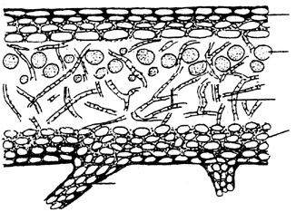
Rhizine
Medulla
Lower cortex
Fig : Transverse section of a foliose lichen
Lower cortex : Comprising of closely packed dark coloured hyphae Rhizoids arise from this layer.
(4) Special structures and propagules : The following specialized structures and propagules are associated with lichen thalli :
- Breathing pores : The upper surface of some lichens, particularly the foliose lichens is provided with pores. Here the fungal hyphae are loosely arranged. They help in aeration.
- Cyphellae : These are small, almost circular depressions present on the lower side of the thallus. The medullary hypae are not exposed through these depressions due to the presence of corticating hyphae. They are meant for exchange of gases e.g., Sticta. Similar structures without any cortical border are called pseudocyphellae.
- Cephalodia : These are gall like outgrowths present on the upper surface of the thallus. They are distinguishable into cortex and medulla with similar fungal but different algal components from that of the main thallus. Lichens having two algal and one fungal partner are called diphycophilous. The cephaloida are meant for retaining moisture e.g, Peltigera.
- Isidia : These are coral like, simple or branched outgrowths present on the upper surface of the thallus. They have the same algae and fungal component as that of the main thallus. They help in photosynthesis as also in vegetative propogation e.g., Parmelia, Peltigera.
- Soredia : It is a powdery mass comprising both algal and fungal components formed in a postule like structure called soralium. The soralia arise from the algal zone lying just below the upper cortex e.g., Physia, Parmelia.
(5) Reproduction : Lichens reproduce both by asexual and sexual methods.
(i) Asexual reproduction
- Fragmentation : The main thallus breaks into small pieces which grow as independent thalli.
- Rejuvination : Plants like Cladonia show this unique phenomenon. It becomes young again. The older parts of the thallus die whereas the young branches continue to grow.
- Isidia : These are small superficial outgrowths on the surface of lichen thallus. They enclose the same alga as present in the thallus and covered by continuous cortex. Their function is to increase the photosynthesis by increasing the surface area. They get detached from the thallus, disseminate by wind and grow into new thalli.
- Soredia : This is a powdery mass formed in a postule like structure called soralium. Each soredium forms a new thallus under favourable conditions.
- Conidia : In serveral lichens the fungal component forms conidia on conidiophores. They form new fungal mycelium which with suitable algal component form the lichen.
- Pycniospores : The conidia formed in a flask shaped structures lying embedded in the thallus (pycnidia) are called as pycniospores. The pycniospores form new fungal mycelium which consitute the lichen on coming in contact with suitable algal component.
(ii) Sexual reproduction : Sexual reproduction in lichen is performed only or mainly by its fungal component. So, the structure of the reproductive organs is dependent upon the type of their fungal partner.
The ascolichens reproduce sexually by forming sex organs. The female sex organ is called ascogonium and the male, pycnidium. The ascogonium is a multicelled structure coiled in its basal region. The terminal region is some what eract and called trichogyne. The ascogonium remains embedded in the thallus. The pycnidia acting as male sex organs are called spermogonia. They are pitcher shaped structures that lie embedded in the thallus. The conidia formed in the spermogonia act as spermatia. Some sterile hyphae also emerge out of the ostiole. The spermatia are colourless tiny structures of varying shapes, they are disseminated by wind. Finally, they attach themselves to the sticky tip of the trichogyne. This is followed by plasmogamy and migration of the male nucleus to the female structure. Ascogenous hyphae now develop from the fertilized cell of ascogonium. The asci develop by crozier formation and karyogamy occurs inside the ascus mother cell. This is followed by meiosis and mitosis resulting in the formation of eight ascospores inside the ascus. Simultaneously, the surrounding hyphae also develop and as a result fruiting body called ascocarp or ascomata is formed. The ascocarp may be an apothecium or perithecium.
Accordingly, the ascolichens are divided into two groups namely gymnocarpae and pyrenocarpae respectively. While the apothecia are cup- shaped structures e.g., Physcia, the perithecia are pitcher shaped e.g., Acrocordia. The fruiting body is internally distinguishable into three zones.
- Thecium : It is the fertile zone comprising fertile asci Asci Paraphyses
| cortex | |
| whereas the thalline margin includes the algal component also. | Fig : V.S. of lichen thallus through apothecia, (A) Lecanorine type, (B) Lecideine type |
Epithecium and sterile paraphyses.
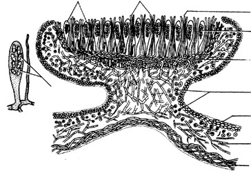
(
A)
(
B)
Ascospore
Thecium
- Epithecium : It is the zone formed by the tips of
Hypothecium paraphyses projecting beyond the asci.
- Hypothecium : It is the zone formed by loosely Algal layer
packed hyphae lying below the thecium. Upper cortex
An apothecium has two types of margins, proper and Medulla thalline. The proper margin is formed by fungal hyphae only Lower
Accordingly, we differentiate two types of apothecia.
- Lecideine type : They have only the proper margin e.g., Lecidea.
- Lecanorine type :They have both proper as well as thalline margin e.g., Lecanora.
The sterile tissue lying in between the asci is sometimes called hamathecium. The asci dehisce releasing the ascospores. The ascospores germinate to form the fungal hyphae. On coming in contact with the suitable algal component, they constitute the lichen. (6) Economic importance
- Pioneer of vegetation : Lichens are considered as pioneers in initiating a plant succession on rocks. These are the first plant group which play an important role in the formation of the soil. So lichens are called as formers of nature or soil builders. Crustose being the first followed by foliose and finally fructicose lichens.
- Food and Fodder : Reindeer moss (Cladonia rangiferina) of the arctic region is the used as food for reindeer and cattle. Iceland moss (Cetraria icelandica) is ground up and mixed with wheat and made into cakes in Iceland. Rock tripe (Umbillicaria) has been eaten by travellers when they face starvation in actic regions. Evernia is used by Egyptians for making bread and Umbilicaria esculenta is regarded as a delicacy in Japan. Species of Parmelia are used as curry powder in India.
- Medicinal uses : Dog lichen (Peltigera canina) was used as medicine for hydrophobia in ancient days and Lungwort (Lobaria pulmonoria) was used for the diseases of lungs. Usnic acid obtained from Usnea (old man’s beard) and Cladonia sp. is used as broad spectrum antibiotic. It is effective against gram positive bacteria. Lobaria pulmonaria, Cetraria icelandica are used in respirotory diseases particularly T.B., Roccella montagnei in angina, Peltigera canina in hydrophobia, Parmelia sexatilis in eipilepsy and Usnea barbata in urinary troubles.
- In perfumery : Ramalina and Evernia, having sweet scented thalli, are used in the preparation of Dhup, Havan Samagri and soap. Perfumes are extracted from Evernia prunastri and Lobaria pulmonaria.
- In tanning and dying : Lichens like Cetraria icelandica and Labaria pulmonaria are used in tanning. A red dye is obtained from Ochrolechia sp. whereas Parmelia sp. yield a brown dye. Litmus used as acid-base indicator is obtained from Roccella montagnei and Lasallia pustulata. An orchill dye is obtained from Roccella and Leconara which is purified as orcein and used as a biological stain.
- In brewing and distilling : The lichens contain carbohydrates in the form of lichenin. Cetraria islandica and Cladonia rangiferina (yield upto 66% of the polysaccharides) are used to obtain alcohol in Sweden and Russia.
- Indicators of air pollution : Lichens are very sensitive to SO2 and grow only in SO2free atmosphere. So lichens like Usnea are used as indicators of air SO2 pollution.
- As poison : Some lichens are poisonous also such as Letharia vulpina due to vulpinic acid, Cetraria juniperina due to pinastrinic acid, Parmelia molliuscula due to selenium, Xanthoria parietina due to beryllium and Everina furfuracea due to chlorine.
- Other uses : Some lichen yield important chemicals e.g., salazinic acid (Ramalina siliquosa), Lecanoric acid (Parmelia subrudecta) and squamatic acid (Cladonia crispata) etc. In hot season, Usnea gets dry and becomes highly inflamable. It easily catches fire and causes forest fires.
| Mycorrhizae. |
The term ‘mycorrhizae’ was coined by Frank (1885) It is an association between a fungus and the root of a higher plant e.g. Pine, Birch, Eucalyptus, Ficus etc. The actively growing roots of higher plants get infected by fungi. As a result, the roots are modified (i.e., become tuberous, nodulated, coralloid, etc.) to accommodate fungi. The root cells and fungi directly transmit nutrient substances to each other. Mycorrhiza is a example of symbiosis or mutualism.
(1) Types of mycorrhizae : Mycorrhizae are classified into three categories :
- Ectotrophic mycorrhiza : It occurs only in about 3% of plant species, majority of which are forest trees, viz. pines, sprues, firs, oaks, beeches, birches, eucalyptus etc. The fungus partner is commonly a basidomycetes. In this type of mycorrhizae, the fungus completely encloses the rootlet in a sheath or mantle of tissue formed of compact hyphal cells and penetrates only between the cells of root cortex. The ectomycorrhizal fungus cannot exist saprotrophically in nature without a plant host association. Such roots are devoid of root hair, root cap and may become unforked, bifurcate, nodular or coralloid.
- Endotrophic mycorrihiza : In this kind of mycorrhizae the fungus does not form an external mantle but lines within the root. The ectomycorrhizae are further divided into three groups :
- Ericaceous mycorrhizae : The fungus forms dense intracellular coils in the outer cortical cells.
- Orchidaceous mycorrhizae : These are associated with orchid roots. The fungus forms association from the time when the orchid seeds germinate.
- Vesicular-arbuscular mycorrhizae (VAM) : The fungi of this group mostly belong to zygomycetes. This type is significant in agriculture because it occurs in a large number of crop plants. The fungal hyphae develop some special organs, called vesicles and arbuscules, within the root cortical cells.
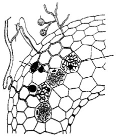
PH
AP
V
SLH
Spore
A
Fig : T.S. of a vesicular-asbuscular mycorrihza (A
Asbuscules, Ap. appresorium, Ph. permanent hyphae, SLH. short-lined hyphas V. Vesicle)
(iii) Ectoendotrophic mycorrhiza : This type of mycorrhiza sharing characteristics of both ecto and endotrophic mycorrhizas. The fungus forms a hyphal mantle and Hartig net as do the ectotrophic mycorrhiza and also establish haustoria and hyphae coils in the epidermal and cortical cells, like the ectorophic mycorrhizas. The external hyphae deliver organic compounds absorbed from the humus to the root cells. One of the best studied examples of ectoendotrophic mycorrhizas is the mycorrhiza of Monotrapa indica, the Indian pipe.
(2) Advantages of mycorrhizal association
- Since all fungi are dependent on some kind of foreign organic matter for their survival, the mycorrhizal fungi obtain their nutrient requirement (primarily simple carbohydrates)from the host plant without damaging the function of root tissues,
- The fungal hyphae increase a plant’s uptake of certain nutrients from the soil, particularly phosphorus, copper, zinc, nitrogen and potassium.
- The mycorrhizal hyphae permeate the soil and help the absorption of water by host more efficiently,
- The mycorrhizal plants need less fertilizer and can even grow better on the infertile soils. They withstand high doses of heavy metals and acid rain pollution.
- The fungi produce various growth promoting substances which help the plants to grow better.
- Due to mycorrhizal association, the higher plants develop resistance to soil borne diseases (due to phytolaxins released by fungi), drought resistance and tolerate salinity, pH and temperature extreams.
Leave a Reply